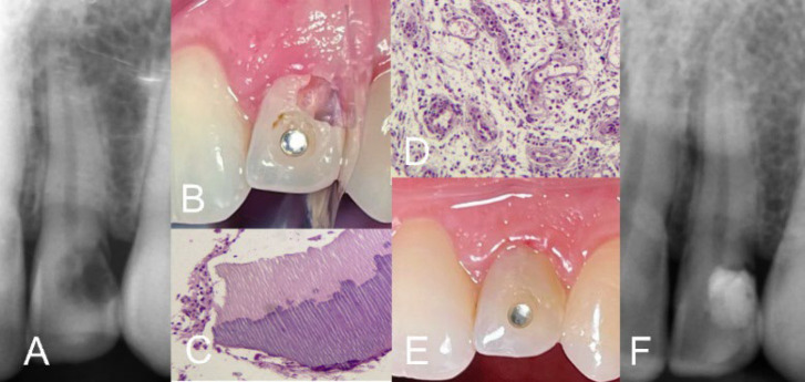
Figure 5.
External Repair of ICRR Class 2 in Upper Left Lateral Incisor. A) Periapical radiograph reveals an ICRR lesion in the involved incisor; B) Non-surgical approach; clinical view after removal of the resorptive tissue from the lesion, exposing the pulp; C and D) Histopathological views of the removed tissue specimen with H&E staining display resorptive dentine pieces with dentinal tubules, and an inflamed tissue; E) Clinical image post-VPT using calcium-enriched mixture and composite filling of the cavity; F) Immediate postoperative radiograph demonstrates the success of VPT and proper coronal filling

