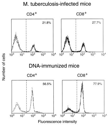
FIG. 2.
Expression of CD44 on CD4+/CD8− and CD8+/CD4− splenocytes when the cells had been amplified by 7-day culture with J774-hsp65 after purification from hsp65 DNA-immunized or M. tuberculosis-infected mice; the cells were stained with MAb against CD44 and analyzed by FACScan. The percentage of cells showing CD44hi fluorescence is shown in the upper right corner of each panel.

