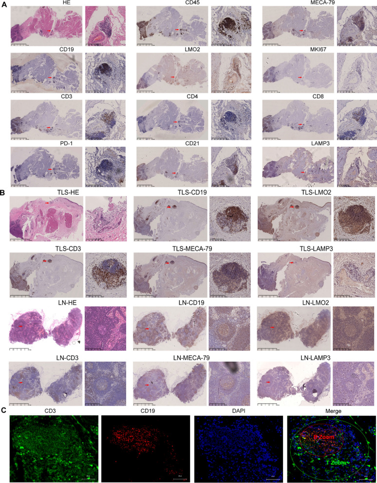
Fig. 5.
TLSs in MIBC. A HE staining shows TLSs (red squares) in MIBC, and IHC staining shows the following markers of TLSs (red squares): CD45, CD19, CD3, MKI67, CD4, CD8, PD-1, CD21, LAMP3, MECA79A and LMO2. Scale bar: 2.5 mm in panoramic images and 100 μm in magnified images. B H&E staining and IHC for the indicated markers for representative LNs (two columns below) and TLSs (two columns above). Scale bar: 2.5 mm in panoramic images and 100 μm in magnified images. C mIF for the indicated markers for representative TLSs. The following markers are shown in individual and merged channels: CD3 (green), T-cell marker; CD19 (red), B-cell marker; DAPI (blue), nuclear marker. Scale bar: 25 μm

