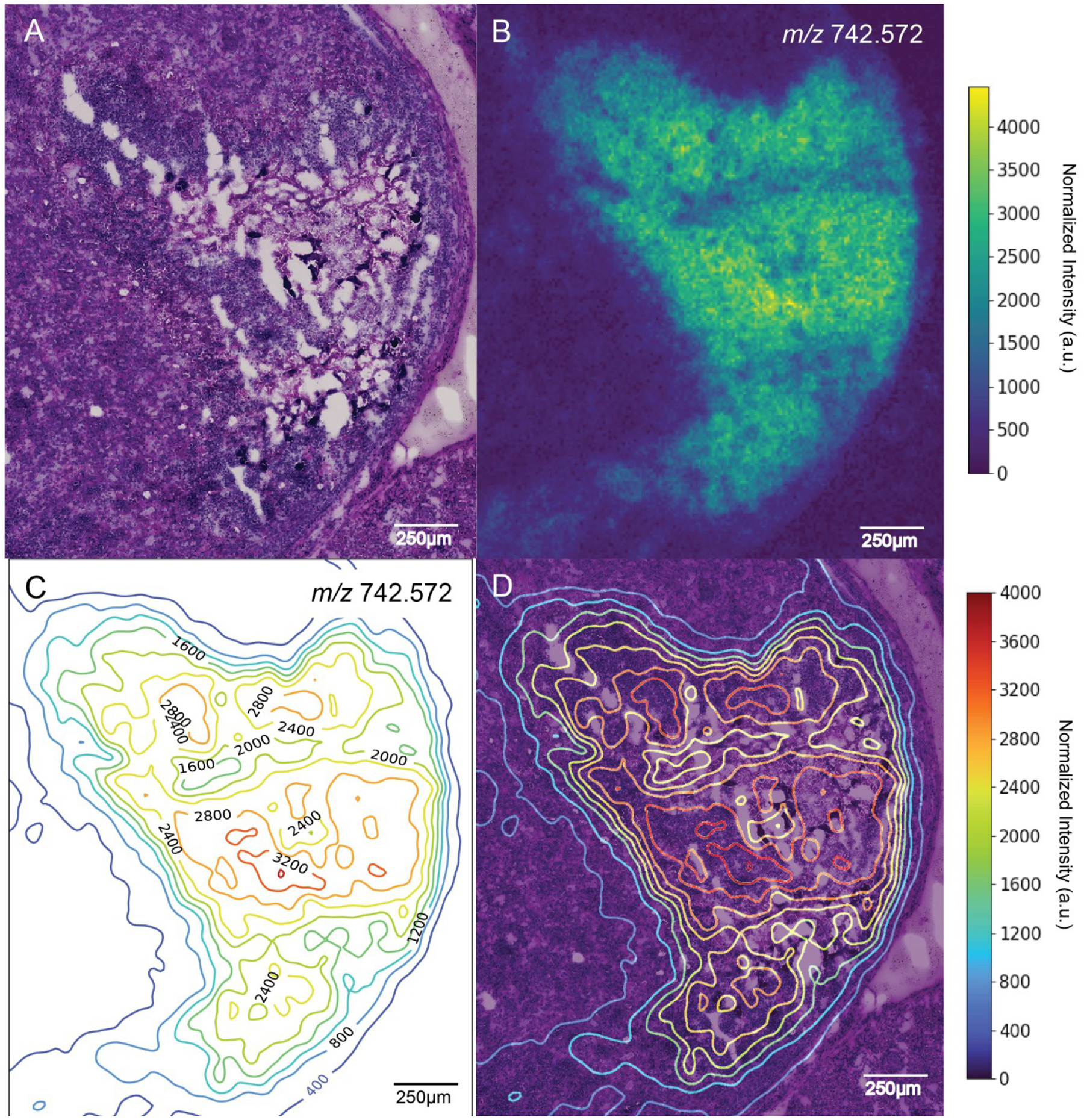
Figure 2.

Contour map of a single MALDI IMS ion image correlating to a staphylococcal abscess. (A) Periodic acid-Schiff (PAS) stain of an S. aureus abscess within the renal cortex of a murine kidney. White areas in the tissue show freeze artifacts resulting from tears. (B) Image of [PC(O-32:0) + Na]+ (m/z 742.572, −0.09 ppm) that was found to colocalize with regions of staphylococcal infection based on a pixelwise Pearson correlation analysis. (C) Contour map generated using the ion image for [PC(O-32:0) + Na]+, with contours labeled by m/z intensity values. (D) Contour map overlaid with PAS.
