
Figure 2.
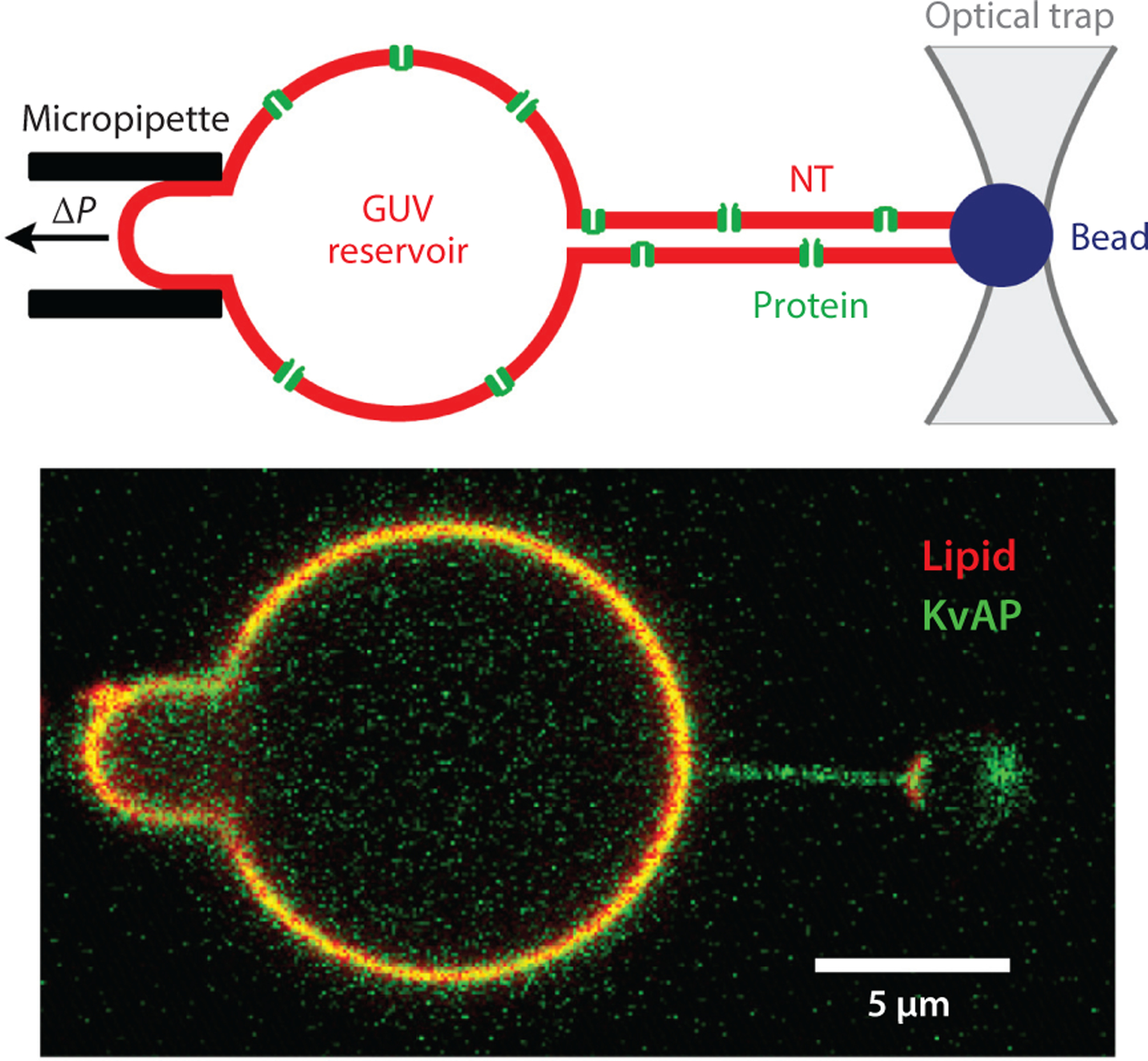
Membrane NTs connected to a reservoir GUV are widely used for quantitative analysis of CCC. (Top) The schematic shows an NT pulled from a GUV reservoir containing transmembrane protein (voltage-dependent K+ channel KvAP) (2). The GUV is held by a micropipette with negative hydrostatic pressure inside. The NT is pulled from the GUV by a microbead controlled by optical tweezers. (Bottom) The fluorescence microscopy image shows the membrane fluorescence coming from a lipid probe (red) and fluorescently labeled KvAP (green). Note that the green fluorescence prevails in the NT, indicating curvature-driven accumulation (sorting) of KvAP in the NT. Figure adapted with permission from Reference 2. Abbreviations: CCC, curvature-composition coupling; GUV, giant unilamellar vesicle; NT, nanotube.