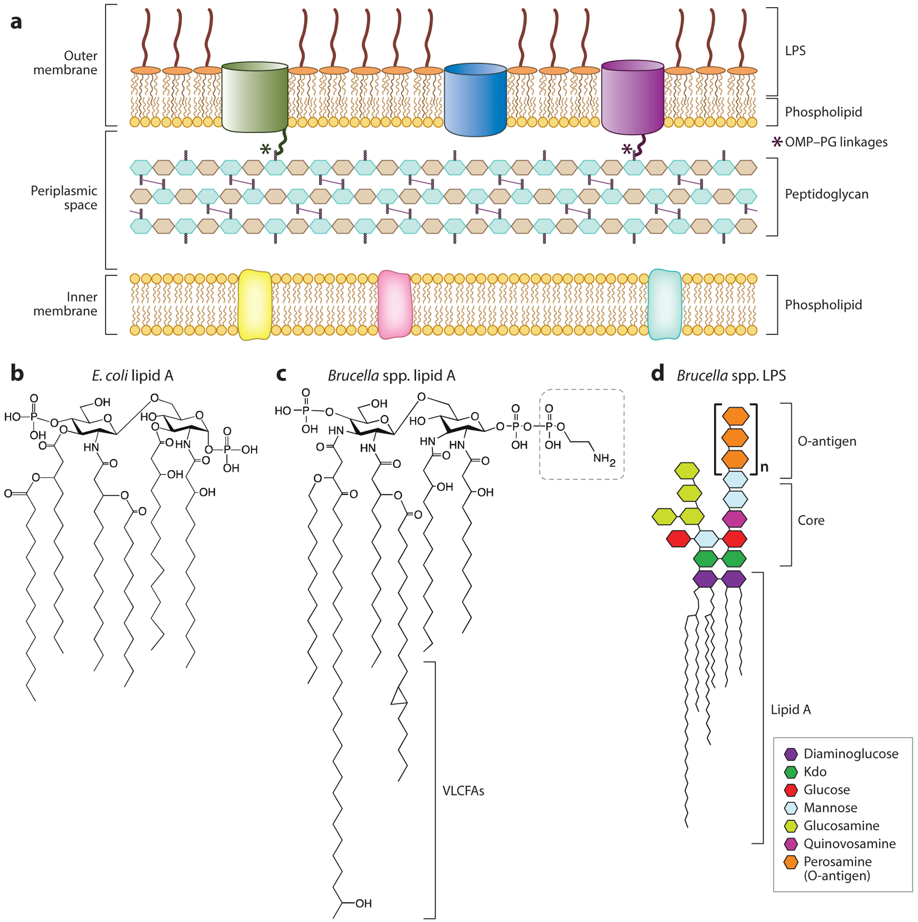
Figure 3.

(a) Overview of the envelope layers of Brucella. Notably, a subset of Brucella outer membrane proteins are covalently linked to the peptidoglycan cell wall via a conserved sequence at the protein N terminus. (b) Chemical structure of Escherichia coli lipid A (100). (c) Chemical structure of Brucella spp. lipid A showing VLCFA tails and pyrophosphorylethanolamine modification of diaminoglucose backbone (outlined with dashed-line box). Structure adapted from model and data presented in References 20 and 29. (d) Brucella smooth LPS structure showing lipid A, core oligosaccharide, and O-polysaccharide (O-antigen), based on models and data presented in References 41, 72, and 117. Abbreviations: Kdo, 3-deoxy-d-manno-2-octulosonic acid; LPS, lipopolysaccharide; OMP, outer membrane protein; PG, peptidoglycan; VLCFA, very-long-chain fatty acid.
