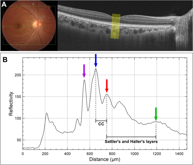
Fig. 1.
Determining choroidal reflectivity. A Choroidal reflectivity was determined on horizontal B-scan images of the swept-source OCT using a line plot of ImageJ with a width of 500 µm. The 500-µm-wide line plot was set perpendicular to Bruch’s membrane (blue arrow). B Using the “plot profile” function of ImageJ, the reflectivity of the choroid was determined. The reflectivity profile plot shows several inflection points, with peak reflectance corresponding to the ellipsoid zone (purple arrow), retinal pigment epithelium (blue arrow), and the choroidal–scleral junction (green arrow). The hyperreflective layer of the inner choroid (red arrow), which had the highest reflectivity within areas with inverted choroidal reflectivity, was observed between the choriocapillaris (CC) and medium-sized and large choroid vessel in Sattler’s and Haller’s layers. CC choriocapillaris, OCT optical coherence tomography

