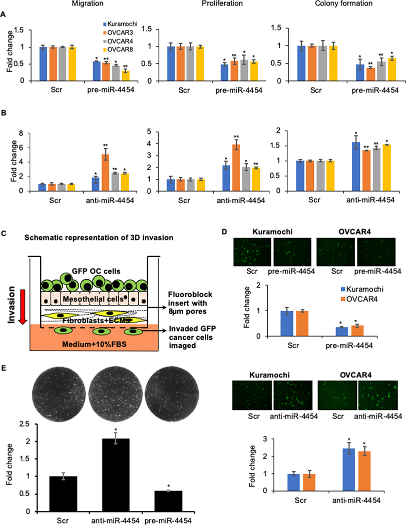
Figure 2.
(A) Overexpression of miR-4454 inhibits motility and growth of OC cells. Kuramochi, OVCAR3, OVCAR4 and OVCAR8 cells were transfected with pre-miR-4454 or scrambled control oligos (Scr) and used for the functional assays. Migration: Transfected cells were seeded in transwell inserts with 8 μm pores and allowed to migrate towards medium containing 10% FBS. Migrated cells were fixed at the optimal time points, imaged and quantified using image-J software (mean ± SD; 3 independents experiments). Proliferation: Transfected cells were seeded in 96-well plates and allowed to grow for 5 days. Cell proliferation was determined by MTT assay (mean ± SD; 3 independents experiments). Colony formation: Transfected cells were seeded in 6-well plates for a colony formation assay. Once visible colonies were formed, the colonies were fixed, stained, imaged and counted using image-J software (mean ± SD; 3 independents experiments). (B) Inhibition of miR-4454 increases the motility and growth of metastatic OC cells. Kuramochi, OVCAR3, OVCAR4 and OVCAR8 cells were transfected with anti-miR-4454 or scrambled control oligos (Scr) and used for the migration, proliferation and colony formation assays as described above (mean ± SD; 3 independents experiments). * p<0.01, ** p<0.05, Students t-test. (C) Invasion through the 3D omentum culture. Schematic representation of the 3D invasion model mimicking the invasion of metastasizing OC cells through the outer layers of the omentum. The 3D omentum culture was assembled in fluoroblock transwell inserts with 8 μm pores. GFP labeled OC cells were seeded on the 3D culture and allowed to invade though it towards the lower chamber containing medium with 10% FBS. (D) Top: Kuramochi and OVCAR4 cells were transfected with pre-miR-4454 or scrambled control oligo (Scr) and evaluated for their ability to invade through the 3D omental culture. Invasion was stopped at the optimal time. The invaded GFP labeled OC cells were imaged using the EVOS FL auto microscope and quantified using image-J software (mean ± SD; 3 independent experiments). Bottom: Kuramochi and OVCAR4 cells were transfected with anti-miR-4454 or scrambled control oligo (Scr) and their ability to invade through the 3D omental culture was evaluated. The invaded GFP cells were imaged and quantified using image-J software (mean ± SD; 3 independent experiments). Representative images of OC cells that have invaded through the 3D omentum culture, for each of these treatment groups are shown above respective graphs. (E). OVCAR8 GFP cells transfected with scrambled control oligos, anti-miR-193b or pre-miR-193b, were seeded on 3D omental culture and allowed to form colonies. The fluorescent colonies were imaged on a Syngene G-Box imaging system and counted using image-J software (mean ± SD; 3 independents experiments). * p<0.01, Students t-test compared to control.

