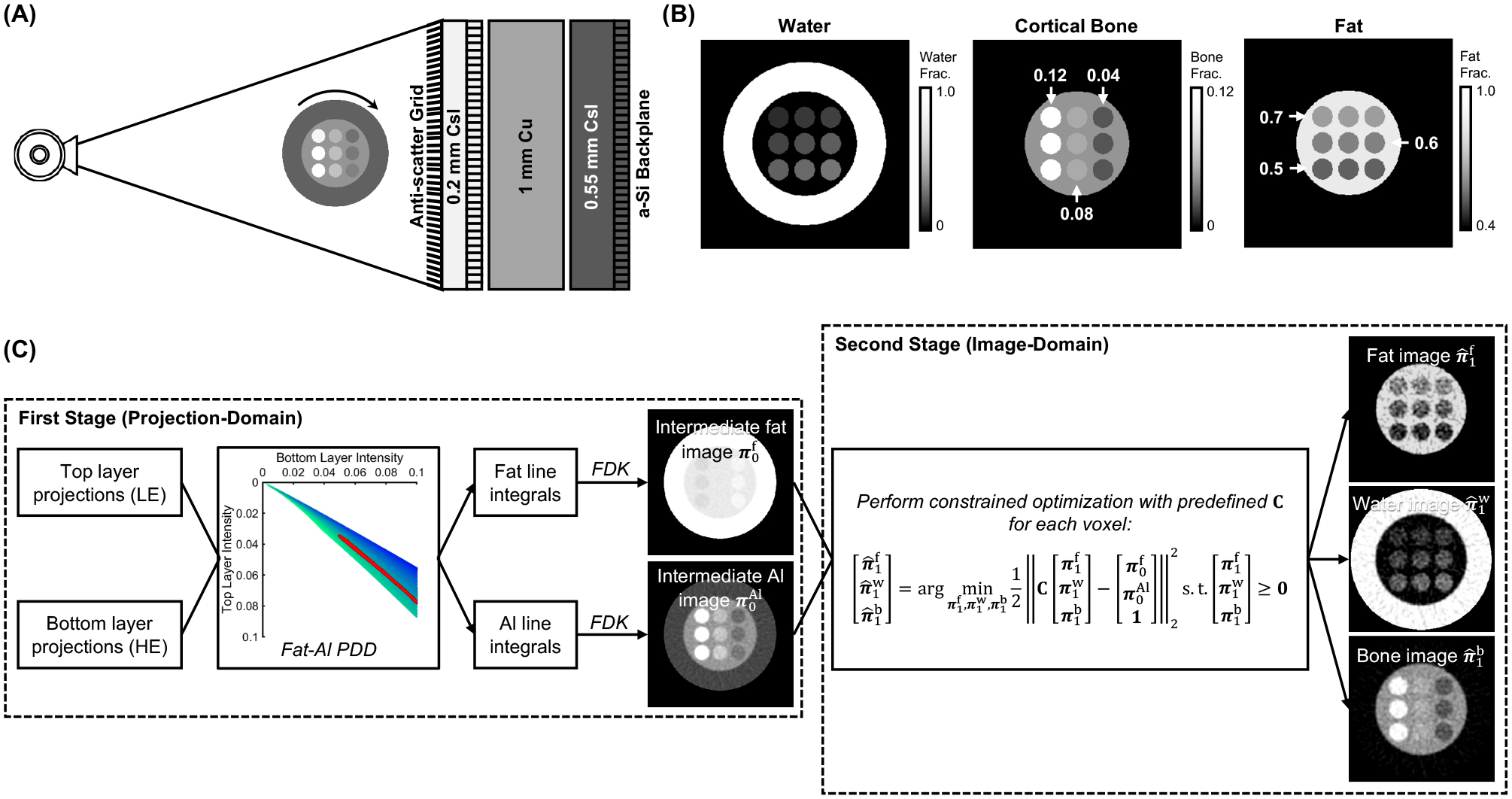
Figure 1.

(A) Imaging configuration for DE CBCT of BME using dual-layer FPD. (B) The three-material phantom (axial views) used for validation. Bone and fat fractions of the inserts are marked on the image, the water fraction was given by the volume conservation principle. (B) The two-stage three-material DE decomposition pipeline.
