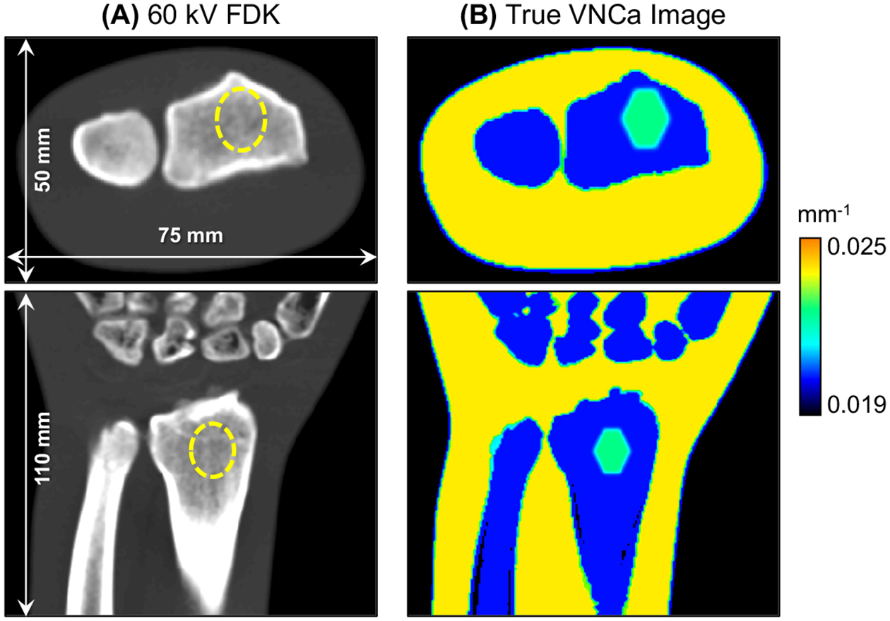
Figure 2.

(A) 60 kV FDK reconstruction and (B) VNCa image of the realistic wrist phantom. The dashed line identifies the location of the BME stimulus in the FDK image. The BME lesion is easily detected as a region of increased VNCa attenuation within the bone marrow of the radius.
