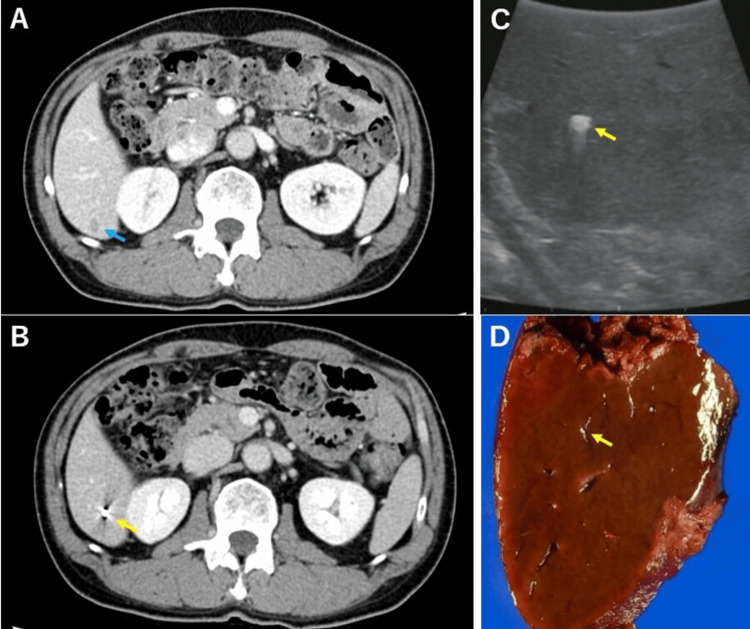
Figure 2. Images from a patient with percutaneous marking.
(A) CT before chemotherapy shows a liver tumor (blue arrow). (B) CT before chemotherapy shows the marking coil (yellow arrow). (C) The marking coil can be confirmed by intraoperative echo, and no tumor can be confirmed (yellow arrow). (D) The marking coil can be confirmed in the excised specimen (yellow arrow).
CT: computed tomography

