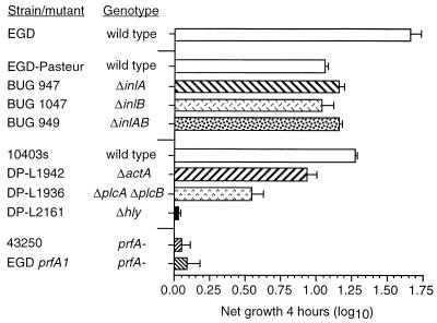
FIG. 1.
Growth of wild-type L. monocytogenes and L. monocytogenes mutants within HUVEC. Confluent HUVEC monolayers were infected with 104 CFU. Cells and bacteria were cocultured for 60 min, washed, and then incubated for another 60 min in medium containing gentamicin to kill extracellular bacteria. Triplicate wells from one plate were lysed, and intracellular bacterial CFU at time zero were quantified by serial dilution and plating. The second plate was incubated for another 4 h (time plus 4 h), and CFU were quantified as before. Intracellular growth during the 4-h interval was calculated as follows: log10 CFU at time plus 4 h − log10 CFU at time zero. Results are shown as the mean (±SEM) log10 growth from three experiments.

