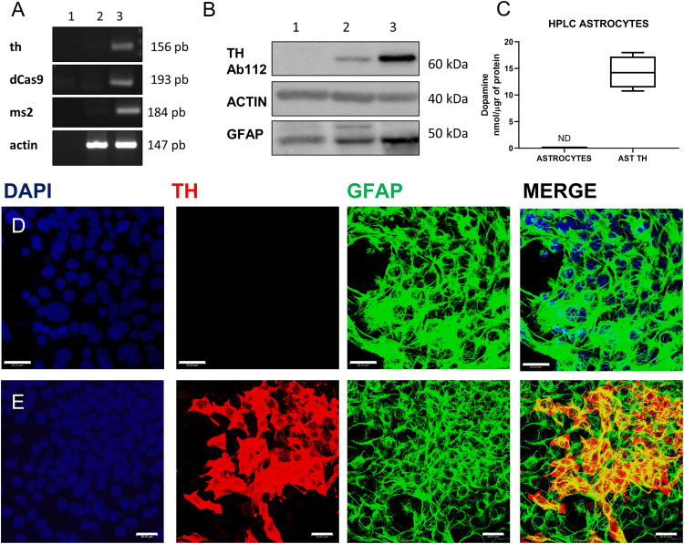
Fig. 2. Expression of Th in astrocytes.
A Detection of th mRNA and the components of the SAM system as determined by RT-PCR in astrocytes; lane 1- H2O (negative control), lane 2- astrocytes, lane 3- astrocyte-TH cells. B Detection of Th protein as determined by Western blot analysis; lane 1- astrocytes, lane 2- astrocyte TH, and lane 3- rat striatum. In A and B, actin is a positive control. C Determination of DA in the extracellular medium, as determined by HPLC in astrocytes and astrocyte-TH cells; independent t-test p = 0.01, n = 3. D Immunofluorescence in astrocytes for Th, GFAP, and DAPI (Nuclei). E Immunofluorescence for Th, GFAP, and DAPI in astrocyte-TH cells. Images in D and E were obtained with confocal microscopy. Bars represent 30 µm.

