Fig. 2.
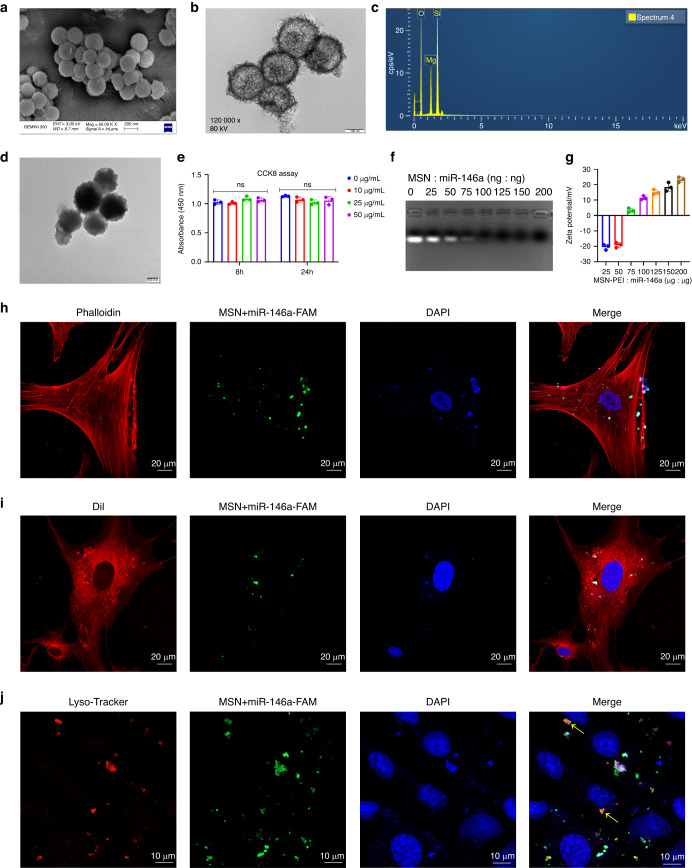
Preparation and characterization of the MSN+miR-146a complex. a–c ECM and TEM images and EDS of MSNs. d TEM images of MSN-PEI. e CCK-8 assay of hDPSCs after 8 and 24 h of coculture with PEI-modified MSNs at various concentrations. f, g Gel retardation and zeta potential tests of the MSN+miR-146a complex at different weight ratios. h–j Cellular uptake assay of MSN+miR-146a-FAM in hDPSCs after 24 h of coculture. Cells were stained to label the cytoskeleton, cell membrane or lysosome. Yellow arrows indicate colocalization of lysosomes and MSN+miR-146a-FAM. ns no significance. One-way ANOVA with Dunnett’s multiple comparisons to the blank group was used

