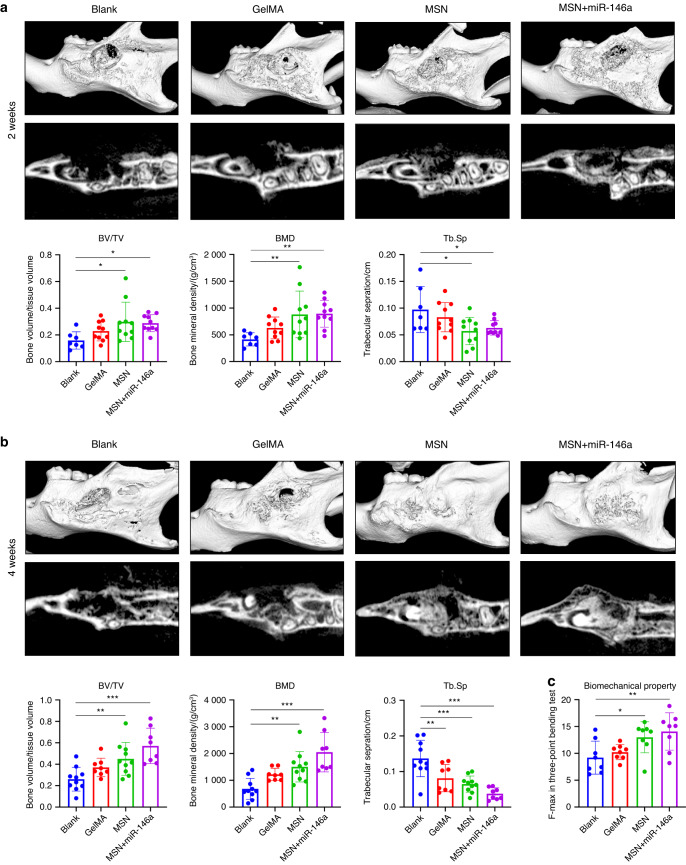
Fig. 5.
MSNs+miR-146a accelerated bone regeneration in a stimulated infected mouse mandibular defect model. a, b Micro-CT examinations of mouse mandible samples after 2 weeks (a) and 4 weeks (b) of healing following surgery with representative reconstructed volume and slice images and the mean BV/TV, BMD and Tb.Sp (n = 7–10). c Maximal force detected in the three-point bending test of trimmed mouse mandible samples (n = 8). *P < 0.05, **P < 0.01, ***P < 0.001. One-way ANOVA with Dunnett’s multiple comparisons to the blank group was used

