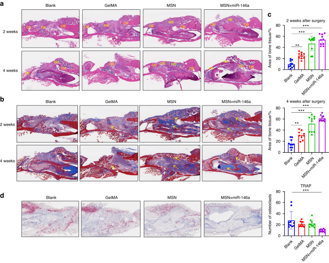
Fig. 6.
MSNs+miR-146a promoted bone regeneration but inhibited osteoclast formation. a HE staining of the mouse mandible samples after 2 and 4 weeks of healing. NB new bone, OB original bone. b Masson staining of the samples after 2 and 4 weeks of healing. Yellow arrows indicate blue-stained type I collagen of new bone. c Statistical analysis of the area proportion of bone tissues in Masson-stained slices of the samples after 2 and 4 weeks of healing (n = 9–10). d TRAP staining and quantitative analysis (n = 10) of the samples after 2 weeks of healing. *P < 0.05, **P < 0.01, ***P < 0.001 One-way ANOVA with Dunnett’s multiple comparisons to the blank group was used

