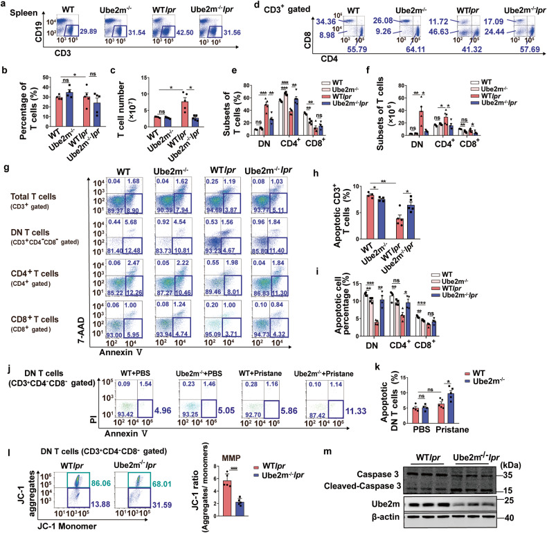
Fig. 4.
Increased DN T cells apoptosis in Ube2m KO lupus-prone mice. a Proportion of T cells in spleens was analyzed by flow cytometry. n = 4 or 5/group. b, c The percentage and number of T cells were calculated based on the data of flow cytometry. n = 4 or 5/group. *P < 0.05. d CD3+ T cell subsets of spleens were analyzed via flow cytometry. n = 4 or 5/group. e, f The percentage and count of T cell subsets were determined using flow cytometry data. n = 4 or 5/group. *P < 0.05, **P < 0.01, ***P < 0.001. g The early apoptosis (Annexin V+/7-AAD-) of total T cells, DN T cells, CD4+ and CD8+ T cells was evaluated with flow cytometry. n = 4 or 5/group. h, i The apoptosis of total T cells and T cell subsets was quantified based on the analysis of flow cytometry. n = 4 or 5/group. *P < 0.05, **P < 0.01, ***P < 0.001. j DN T cell apoptosis (Annexin V+/PI-) in spleens from pristine-induced mice was analyzed with flow cytometry. n = 4 or 5/group. k DN T cell apoptosis in spleens of pristine-induced mice was quantified based on the analysis of flow cytometry. n = 4 or 5/group. *P < 0.05. l MMP was assessed using JC-1 assay to indicate the apoptosis of DN T cells. n = 5/group. ***P < 0.001. m Cleaved-caspase 3 was detected via immunoblotting assay to indicate the apoptosis of DN T cells. n = 3/group

