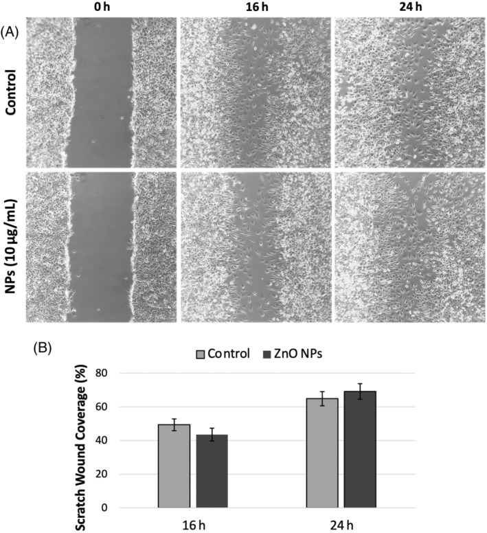
FIGURE 6.

(A) In vitro wound healing assay images of L929 fibroblast cells obtained at 16 and 24 h after wound creation (Co‐ZnO nanoparticles [NPs]‐10 μg/mL). (B) Percentage wound closure graph of L929 fibroblast cells. Results are expressed as mean ± SEM.
