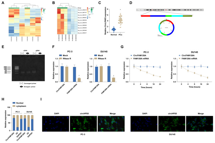
Figure 1.
circFAM126A expression in PCa. A and B: Clustering and scatter plots from circRNA microarray data exhibit the differential expression of circRNAs in PCa groups compared to normal groups, with red indicating high expression levels and green signaling low expression levels. C: The expression of hsa_circ_0001971 in 46 pairs of matched PCa and adjacent non-tumor tissues was detected using RT-qPCR. D: Genomic locus and circular structure of hsa_circ_0001971. E: The presence of hsa_circ_0001971 was validated using divergent and convergent primers for cDNA and gDNA. F and G: RT-qPCR experiments were conducted to assess the effect of RNase R and Actinomycin D on the RNA stability of CircFAM126A and its linear counterpart, FAM126A, in cells. H: The subcellular localization of CircFAM126A was determined through nuclear and cytoplasmic separation experiments. I: FISH experiments revealed that CircFAM126A is predominantly localized in the cytoplasm. Data are expressed as mean ± SD (n = 3), with * P < 0.05 indicating statistical significance.

