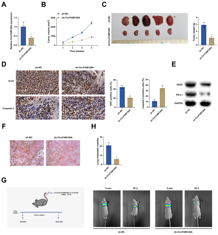
Figure 9.
Silencing circFAM126A inhibits PCa xenograft tumor formation. A: The expression of CircFAM126A in tumors was investigated using RT-qPCR. B and C: Measurements were taken for both the volume and weight of the tumors to assess their growth. D: Immunohistochemical staining techniques were applied to detect the cellular proliferation marker Ki67 and the activated apoptosis indicator cleaved caspase-3 in tumor tissues. E: The protein levels of VEGF and PD-L1 in tumor samples were examined using Western blot analysis. F: Oil Red O staining was utilized to quantify the lipid content present in lung tissues. G: Bioluminescence imaging provided a visualization of the metastatic spread of cancer cells to the lungs. H: A quantitative assessment was made regarding the lymph nodes within lung tissues. Data are presented as mean ± SD (n = 6), with * P < 0.05 indicating statistical significance.

