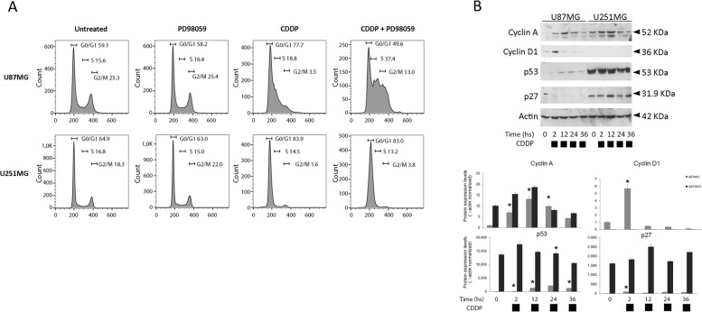
Fig. 2.
Cisplatin-based chemotherapy induces cell-cycle arrest in human glioblastoma cells. Panel A—Flow cytometric DNA content (propidium iodide, FL2 fluorescence) analysis in cells following FLH2 versus FLW2 analysis for doublet elimination. U87MG and U251MG were treated with single-dose cisplatin (CDDP) 16,6 µM and/or PD98059 (40 μM). Values are expressed as mean ± s.e.m. (n = 3). Differences between treatments were tested for statistical significance using Student’s matched pairs t-test (*P < 0.0001 compared to untreated sample). Panel B – Representative immunoblots showing Cyclin-A, Cyclin D1, p53 and p27 protein levels in U87MG and U251MG cells treated with cisplatin (CDDP) 16,6 µM for 2, 12, 24 and 36 h. Western blot analysis of β-actin was performed in each experiment, as loading control. Histograms below reports the relative expression of proteins normalized to β-actin

