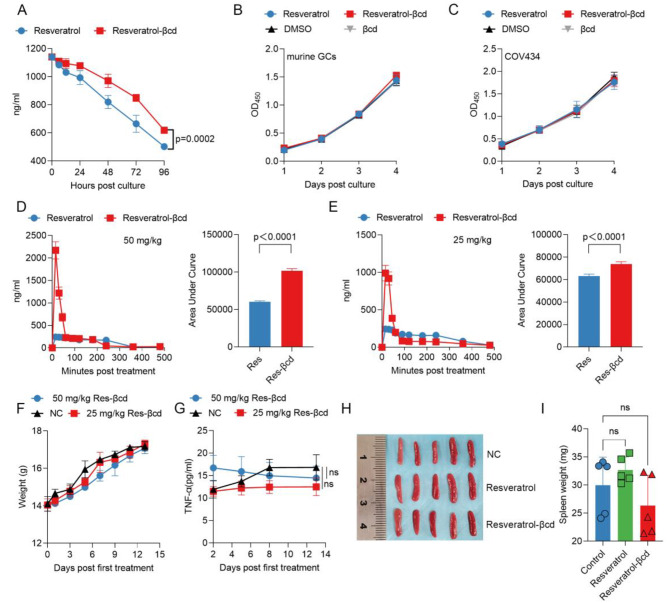
Fig. 1.
Analysis of the effects of resveratrol-βcd on the proliferation of ovarian granulosa cells and the immune response. (A) 6 ml DMEM culture medium was added to a 25 cm2 cell culture flask. For different groups, 5 µM (1141.2 ng/ml) resveratrol-βcd or resveratrol was added. 200 µl of supernatant at 6, 12, 24, 48, 72, and 96 h after incubation was collected, and the resveratrol concentration in the culture medium was detected using ELISA. n = 3; (B) Primary mouse granulosa cells or (C) COV434 cells were cultured with 5 µM resveratrol-βcd or resveratrol. The culture medium was replaced every 24 h to maintain the drug concentration. On days 1, 2, 3, and 4 after drug treatment, a CCK-8 assay kit was used to detect cell proliferation. n = 3; the statistical results are not significant among groups. (D) Resveratrol-βcd or resveratrol was administered by gavage at a dose of 50 mg/kg or (E) 25 mg/kg, and the levels of resveratrol in the blood were measured at corresponding time points after gavage. The statistical results of the AUC are on the right side of each line graph, n = 5; (F) Resveratrol-βcd or resveratrol was administered by gavage at a dose of 25 mg/kg or 50 mg/kg every other day, and the changes in body weight of the mice during the treatment period are shown, n = 5; the statistical results are not significant among groups. (G) The changes of TNFα in the serum during the treatment period. n = 5; (H) Typical images of mouse spleens on day 13 after drug administration and (I) their weights, n = 5. Each group used 5 mice for experiment, in vivo experiments in this study were independently repeated three times. Statistical analysis was performed using two-way ANOVA (A,B,C,F,G), and unpaired two-tailed Student’s t test (D,E). one-way ANOVA (I), comparison was made between control vs. resveratrol-βcd, control vs. resveratrol; ns, not significant

