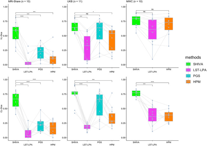
FIGURE 4.

Voxel (VL‐) and cluster‐level (CL‐) Dice scores of SHIVA‐white matter hyperintensity (WMH) compared with lesion prediction algorithm implemented in the lesion segmentation toolbox (LST‐LPA), PGS, and HyperMapper (HPM) tools in the test‐set subjects in each cohort. Comparisons of VL‐Dice (top row) and CL‐Dice (bottom row) scores between SHIVA‐WMH against the reference tools (LST‐LPA, PGS, and HPM) are shown, separately for MRi‐Share (n = 10), UKB (n = 11), and MWC (n = 10) test subjects. Asterisk indicates the degree of statistical significance for each paired t test comparing SHIVA‐WMH against each of the reference methods: ****p < .0001, ***.0001 ⩽ p < .001, **.001 ⩽ p < .01, *.01 ⩽ p < .05, ns p ⩾ .05.
