
Figure 5. Collagen denaturation associated with pathological remodeling revealed via collagen hybridization.
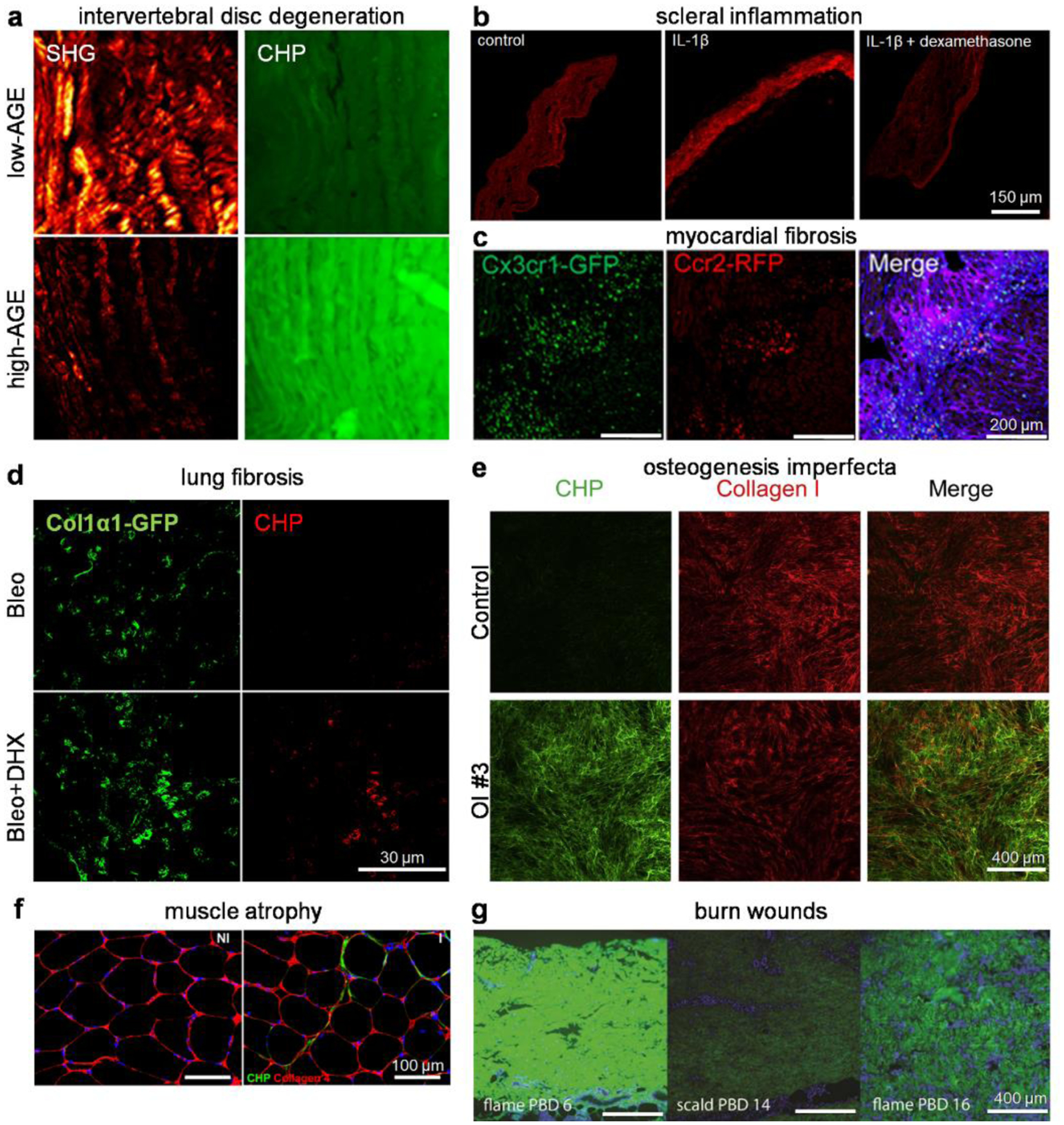
(a) High advanced glycation end product (AGE) diets caused collagen degradation in the central anterior annulus fibrosus of mouse intervertebral disks, as shown by a marked decrease in SHG intensity and an increase in CHP staining. Adapted with permission under a Creative Commons Attribution 4.0 License from ref 135. Copyright 2020 Wiley Periodicals LLC. (b) The IL1-β-induced collagen degradation in organotypic cultured mouse scleral tissue was abolished with dexamethasone treatment. Adapted with permission from ref 139. Copyright 2021 Springer Nature. (c) Histological images of ischemia-reperfusion injured hearts of mice that received a bone marrow mononuclear cell treatment showed localization of CCR2+ and CX3CR1+ macrophages within areas of active collagen remodeling (CHP: purple) in the infarct border zone. Adapted with permission from ref 142. Copyright 2019 Springer Nature. (d) Dihydrexidine (DHX) treatment promoted fibroblast-mediated collagen degradation, evidenced by an enhancement in CHP staining that colocalized with Col1α1-GFP+ fibroblasts in fibrotic lungs of mice treated with DHX in addition to bleomycin. Adapted with permission from ref 144. Copyright 2020 The Company of Biologists Ltd. (e) The ECM produced by cultured dermal fibroblasts isolated from patients with osteogenesis imperfecta (OI) contained a heavy portion of misfolded collagen I, estimated by the fluorescence ratio between the CHP and collagen I stains. Adapted with permission under a Creative Commons Attribution 4.0 License from ref 146. Copyright 2020 Elsevier Inc. (f) Enhanced CHP staining indicative of greater ECM turnover was noted in the injured (I) limb quadriceps muscle compared to the non-injured (NI) ones. Adapted with permission from ref 155. Copyright 2019 SAGE Publications. (g) Collagen denaturation assessed by CHP staining on human burn wound eschar after scalding or flame burn at different post-burn days (PBD). Adapted with permission from ref 156. Copyright 2020 Wound Healing Society.