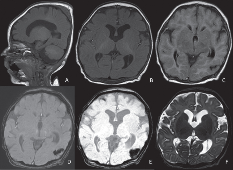
Fig. 2.
Control brain MRI of patient #1 at 3 months of age. Dilated ventricualr system (especially at the lateral ventricles level, A-F), increased CSF spaces in the rolandic areas bilaterally, chronic evolution of a focal haemorrhage at the left temporo-occipital carrefour. A: sagittal T1, B: axial T1, C: axial FLAIR, D: axial susceptibility-weighted phase (SWIp) fast, E: axial minimum intensity projection (miniP), F: axial T2-weighted sequence.

