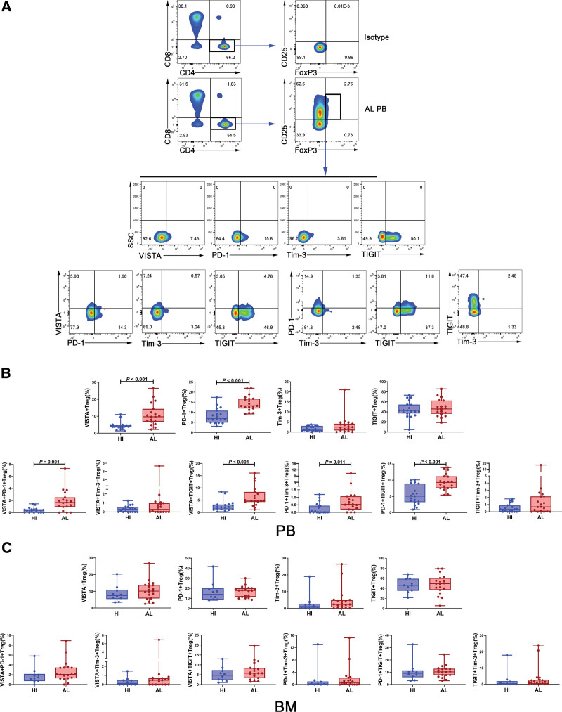
Figure 3.
The percentage of VISTA/PD-1/Tim-3/TIGIT+ Treg in PB and BM from patients with AL amyloidosis and HIs. (A) The analytic logic of flow cytometry detection of single positive (VISTA or PD-1 or Tim-3 or TIGIT) Treg or double-positive (co-expression with each other) Treg in PB or BM. (B) Comparison of the percentage of VISTA/PD-1/Tim-3/TIGIT+ Tregs in PB between patients with AL amyloidosis (n = 19) and HIs (n = 19). (C) Comparison of the percentage of VISTA/PD-1/Tim-3/TIGIT+ Tregs in BM between patients with AL amyloidosis (n = 19) and HIs (n = 10). AL = amyloid light chain, BM = bone marrow, HIs = healthy individuals, PB = peripheral blood, PD-1 = programmed cell death-1, TIGIT = T cell immunoreceptor with Ig and ITIM domains, Tim-3 = T cell immunoglobulin and mucin-domain-containing-3, VISTA = V-domain immunoglobulin suppressor of T cell activation.

