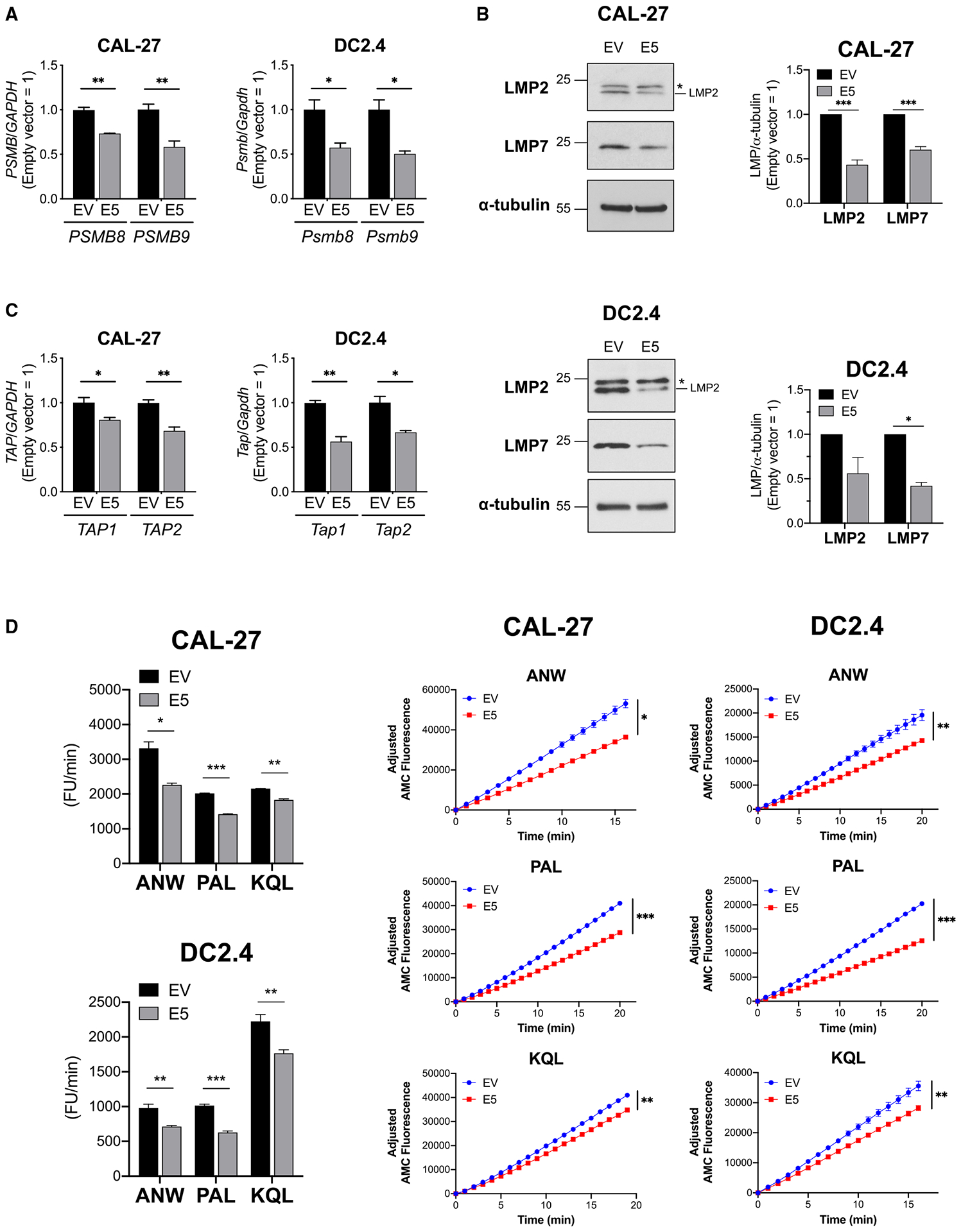
Figure 2. HPV E5 downregulated key antigen-processing molecules, immunoproteasome, and TAP.

(A–C) Transcripts and proteins in HPV E5-expressing CAL-27 and DC2.4 cells were analyzed.
(A) mRNA expression of immunoproteasome subunits analyzed by qPCR.
(B) Western blot of immunoproteasome subunits. An asterisk indicates a non-specific signal.
(C) mRNA expression of TAP analyzed by qPCR.
(D) Immunoproteasome activities in HPV E5-expressing CAL-27 and DC2.4 cells were analyzed using fluorescence-conjugated substrates. Left: average of fluorescent units (FUs) per minute throughout the time monitored. Right: fluorescence kinetics of three substrates. Data are represented as mean ± SEM (n = 3).
