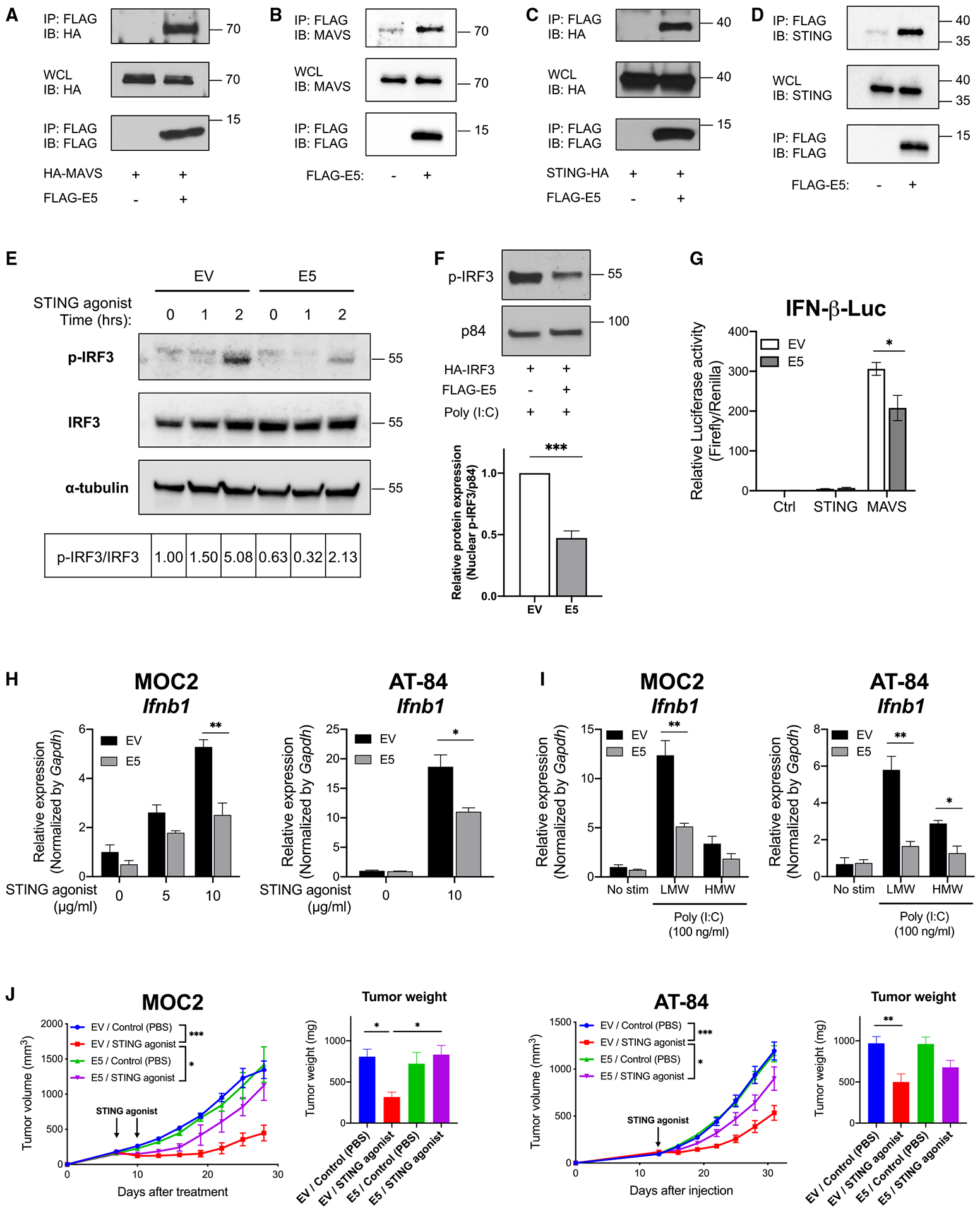
Figure 6. HPV E5 interacted with MAVS and STING and inhibited downstream pathways.

(A) The indicated constructs were transiently transfected in HEK293T cells, and co-immunoprecipitation was performed.
(B) Co-immunoprecipitation of stably expressed E5 and endogenous MAVS was performed with CAL-27.
(C) Indicated constructs were transiently transfected in HEK293T and co-immunoprecipitation was performed.
(D) Co-immunoprecipitation of stably expressed E5 and endogenous STING was performed with CAL-27 cells.
(E) Empty vector- or E5-expressing CAL-27 cells were treated with a STING agonist (10 μg/mL) for 1 or 2 h. Phosphorylation of IRF3 was analyzed by western blotting. The ratio of p-IRF3/total IRF3 is shown.
(F) The indicated constructs were transfected in HEK293T cells. Proteins from nuclear fractions were isolated and analyzed by western blotting (n = 3).
(G) Control, STING, or MAVS constructs were transfected in empty vector- or E5-expressing CAL-27 cells for 24 h. IFN-β promoter activity was measured by a dual-luciferase assay system (n = 3).
(H) mRNA expression after STING agonist treatment was analyzed by qPCR. Cells were treated with a STING agonist for 4 h at the indicated concentrations (n = 3).
(I) mRNA expression was analyzed by qPCR. Cells were treated with 100 ng/mL of poly(I:C) for 4 h (n = 3).
(J) Murine HNSCC lines (left: 1 × 10⁵ MOC2 cells; right: 5 × 10⁵ AT-84 cells) expressing empty vector or E5 were injected into mice (MOC2, n = 4–6 per group; AT-84, n = 6–7 per group). Tumors were treated with a STING agonist (ADU-S100) 20 μg two doses (MOC2) or 20 μg one dose (AT-84) when tumors reached 6–7 mm in diameter. Tumor weight at the time of sacrifice is also shown. Data are represented as mean ± SEM.
