FIGURE 2.

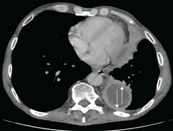
Computed tomography of the chest with a pleural-phase contrast axial cut showing left parietal and visceral pleural enhancement: the split sign (arrows).

Computed tomography of the chest with a pleural-phase contrast axial cut showing left parietal and visceral pleural enhancement: the split sign (arrows).