
Fig. 3.
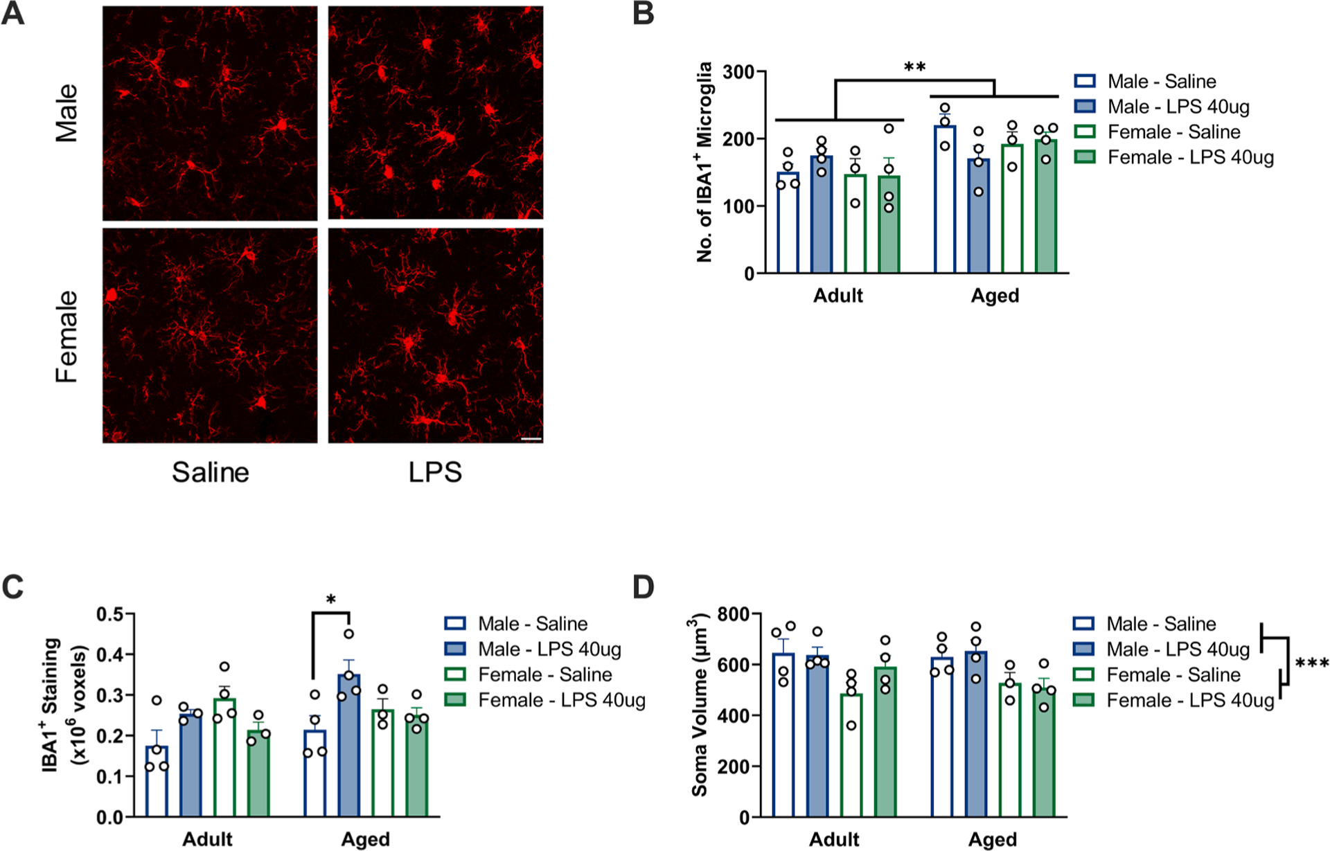
Microglia in aged male, but not aged female, hippocampi show morphological features of inflammatory priming. a) Representative images from the dentate gyrus of aged animals. Scale bar = 20 μm. b) An increased number of IBA1+ microglia was observed in the dentate gyrus of aged animals, ROI: 0.63×0.63 mm, n = 3–4. c) Aged males showed an increase in the area of IBA1+ staining within the dentate gyrus, n = 3–4. d) Males exhibited a larger average soma volume than females in the dentate gyrus, n = 3–4. * p < 0.05, ** p < 0.01, *** p < 0.001.