Fig. 1.
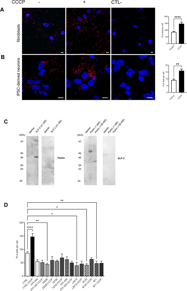
Parkin and SLP-2 interact in human control fibroblasts and hiPSC-derived neurons. A Representative PLA staining for fibroblasts of a control individual under normal culture conditions and after CCCP treatment (3 h, 10 µM). The PLA signal is visualized in red, DAPI-stained nuclei are shown in blue. Scale bar: 10 µm. Quantification of PLA dots per cell shows a significantly higher PLA signal after CCCP exposure, indicating an increased interaction between the two proteins. Statistical differences were calculated by unpaired Mann Whitney U test ****p ≤ 0.0001. B hiPSC-derived neurons of a control individual were processed using PLA under normal culture conditions and after CCCP treatment (3 h, 10 µM). Scale bar: 10 µm. Quantification of PLA dots per cell shows a significantly higher PLA signal after CCCP exposure. Statistical differences were calculated by unpaired Mann Whitney U test. **p ≤ 0.005. The specificity of the PLA interaction results was confirmed by performing the experiments with only one of the two primary antibodies. C Far-Western blot analysis using recombinant SLP-2 (aa 41–356) and Parkin proteins (aa 1–465 and aa 138–465) shows the direct binding of the two proteins. D Quantification of PLA dots per cell in control and PRKN mutant fibroblast lines under normal culture conditions and after CCCP treatment (3 h, 10 µM) using an antibody directed against the Parkin N-terminus and an anti-SLP-2 antibody. Only in the control lines, CCCP treatment resulted in a significantly increased interaction. Statistical differences were calculated by one-way ANOVA followed by Tukey’s post hoc test to correct for multiple comparisons. * ≤ 0.05, **p ≤ 0.01, ****p ≤ 0.0001

