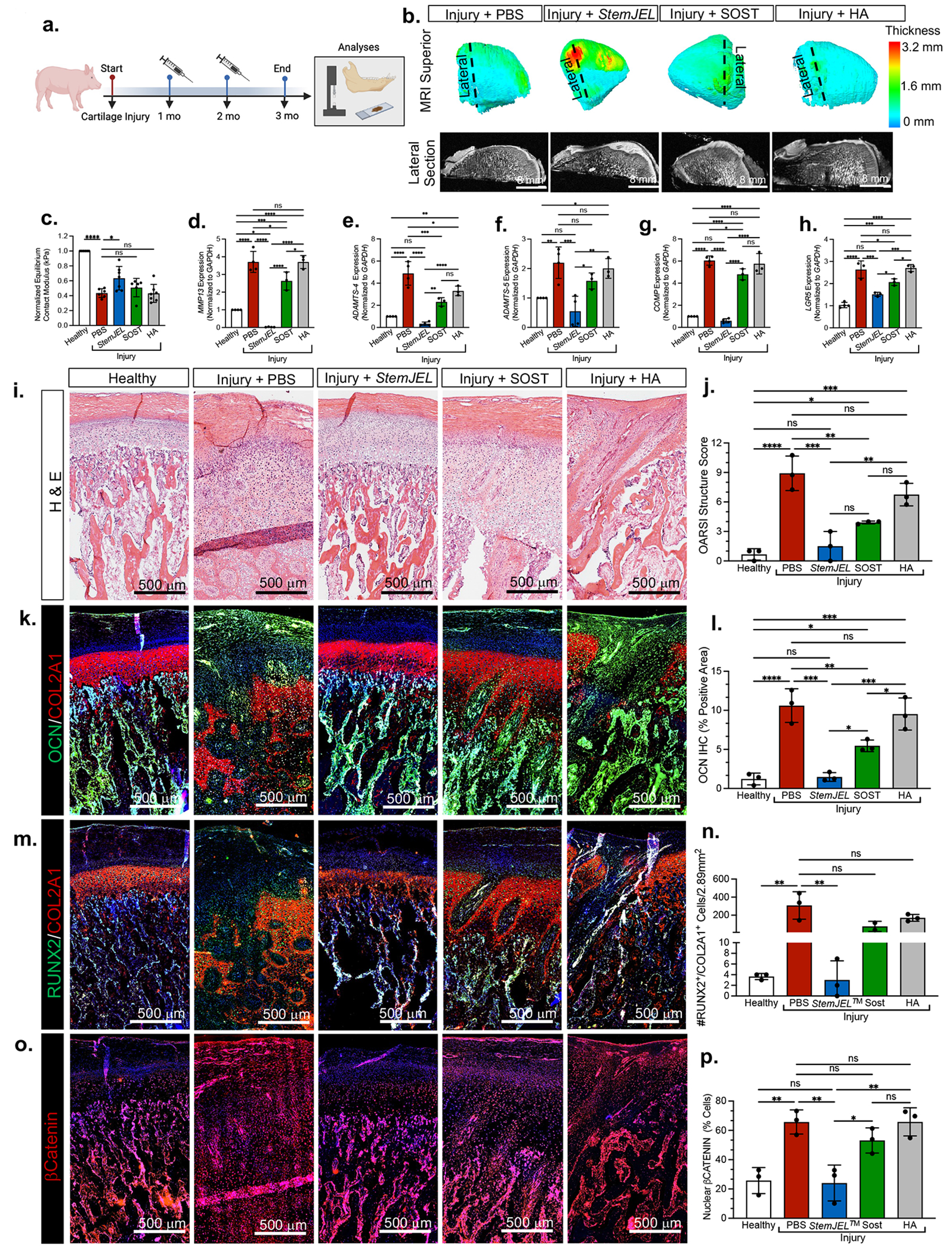
Figure 7. StemJEL™ ameliorates post-traumatic osteoarthritis and restores chondrocyte identity in pre-clinical mini-pig jaw joints.

(a) Schematic of mini-pig jaw joint injury model. (b) MRI of superior view of mini-pig condyles (top). Cross section of MRI (bottom). (c) Normalized equilibrium contact modulus in injured mini-pig condyles normalized to uninjured site within the same condyle. Data are normalized mean ± SD; *p≤0.05, ***p≤0.001; one-way ANOVA followed by Tukey’s post hoc; n=6-8 mini-pigs. (d-h) qRT-PCR using mini-pig mandibular condyles from a. Data are mean normalized to GAPDH ± SD; *p≤0.05, **p≤0.01; ***p≤0.001; ****p≤0.0001; one-way ANOVA followed by Tukey’s post hoc; n=3-4 mini-pigs. (i) H&E staining of mini-pigs from a. (j) OARSI macroscopic score of mini-pig condyles from a. Data are mean score ± SD; *p≤0.05, **p≤0.01; ***p≤0.001; ****p≤0.0001; one-way ANOVA followed by Tukey’s post hoc; n=3 mini-pigs. (k) Immunohistochemistry of osteocalcin (OCN, green) and type II collagen (COL2A1, red) in mini-pig condyles from a. (I) Area of OCN immunostaining in k. Data are mean area ± SD; *p≤0.05, **p≤0.01; ***p≤0.001; ****p≤0.0001; one-way ANOVA followed by Tukey’s post hoc; n=3 mini-pigs. (m) Immunohistochemistry of RUNX2 (green) and type II collagen (COL2A1, red) in mini-pig condyles from a. (n). The percentage of RUNX2+/COL2A1+ cells from immunostaining in m. Data are mean percent ± SD; **p≤0.01; ***p≤0.001; one-way ANOVA followed by Tukey’s post hoc; n=3 mini-pigs. (o) Immunohistochemistry of βCATENIN from mini-pigs in a. (p) The percentage of βCatenin+ cells from o. Data are mean ± SD; **p≤0.01; one-way ANOVA followed by Tukey’s post hoc; n=3 mini-pigs.
