Fig 1.
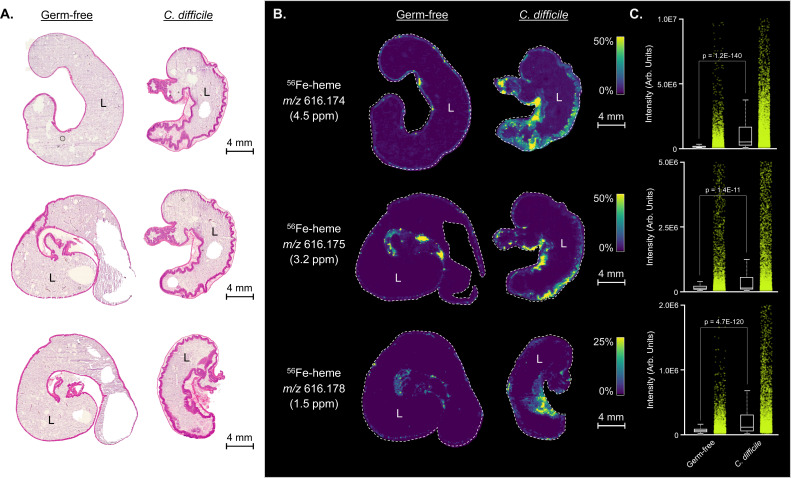
(A) Hematoxylin and eosin-stained cecal tissue sections from germ-free and C. difficile (CD196) mono-infected mice (n = 3 tissue replicates). L marks the lumen of the cecum. (B) Matched MALDI imaging mass spectrometry ion images of 56Fe-heme displaying heme localization to the mucosa of the infected intestinal tract. (C) Relative quantitation of MALDI imaging mass spectrometry 56Fe-heme intensities between germ-free and C. difficile (CD196) mono-infected mice.

