Fig 1.
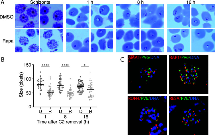
Development of parasites expressing or lacking PV6 during the ring stage and PV6 localization in schizonts. (A) Synchronized parasites with a floxed pv6 (pfa0210c/ PF3D7_0104200) gene were treated at the late trophozoite stage with DMSO or with rapamycin to induce the excision of pv6. Parasites were examined at the schizont stage in Giemsa-stained smears, and subsequent egress was synchronized with ML10 treatment. Giemsa smears were produced 1, 8, and 16 h after Compound 2 (C2) removal. The scale bar represents 5 µm. (B) Quantitation of the size of the parasite diameter in the samples shown in panel A. Data are based on the measurement of at least 30 rings. The Mann–Whitney U test was performed for statistical analysis: *P < 0.05; ****P < 0.0001. D, DMSO-treated; R, rapamycin-treated. (C) Costaining of schizonts with antibodies against PV6 (green) and the apical organelle markers AMA1 (micronemes), RAP1 (rhoptry), RON4 (rhoptry neck), and RESA (dense granule) (all in red) by immunofluorescence microscopy. DNA was stained with Hoechst 33342 (blue). Individual channels are shown in Fig. S1B. The scale bar represents 5 µm.

