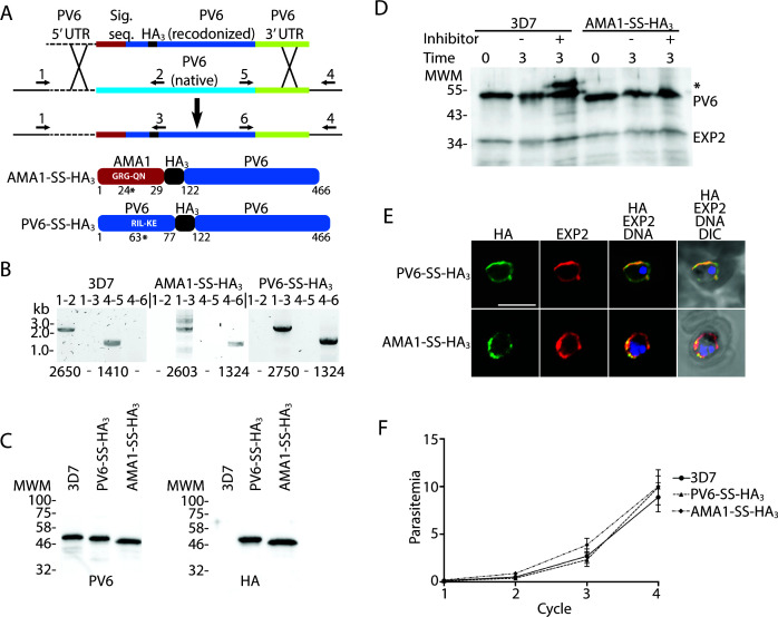
Fig 3.
Replacement of native PV6 with an AMA1-SS-HA3-PV6 fusion. (A) Outline of the replacement strategy of the native pv6 gene with the gene encoding the AMA1-SS-HA3-PV6 fusion. The same strategy was used to create the PV6-HA3-PV6 fusion. Cartoons at the bottom show the expected protein products after the replacement of the native gene with the inserted gene. Indicated in white letters is the predicted signal sequence cleavage site in the AMA1-SS-HA3-PV6 fusion and the PEXEL sequence of the PV6-HA3-PV6 fusion. The number and * indicate the signal sequence cleavage site in the AMA1-SS-HA3-PV6 and PM V cleavage site in the PV6-HA3-PV6 fusion. Numbers indicate amino acid residues of the native proteins. (B) Integration PCR using wild-type and transfected parasite DNA. Primer pairs are indicated above the lanes (see also Table S1), primer binding sites are indicated in panel A, and expected sizes of PCR products are indicated below each lane. Note the absence of integration-specific PCR products in the samples using gDNA from wild-type parasites and the absence of wild-type-specific PCR products in the samples using gDNA from transfectants. (C) Anti-PV6 (left) and anti-HA (right) immunoblots of wild-type (3D7) and transgenic parasite (PV6-HA3 and AMA1-SS-HA3) extracts using the indicated antibodies. Expected molecular weights are 46.4 kDa (3D7 native PV6), 45.4 kDa (PV6-HA3-PV6), and 44.4 kDa (AMA1-SS-HA3-PV6). (D) Effect of Plasmepsin V inhibitor treatment on PV6, AMA1-SS-HA3-PV6, and EXP2. The predicted sizes of uncleaved and cleaved proteins are 53.6 and 46.4 kDa (PV6), 47.3 and 44.4 kDa (AMA1-SS-HA3-PV6), and 33.4 and 30.8 kDa (EXP2). Note the absence of unprocessed AMA1-SS-HA3-PV6 and EXP2 in the presence of the Plasmepsin V inhibitor. * indicates the higher molecular mass product in the 3D7 lysate. (E) Localization of AMA1-SS-HA3-PV6 and PV6-HA3-PV6 fusion proteins. The fusion proteins were detected using an anti-HA antibody, and EXP2 was detected using an anti-EXP2 rabbit anti-serum. (F) Growth assay of 3D7, PV6-HA3-PV6, and AMA1-SS-HA3-PV6 parasites. Individual growth assays were set up in triplicate; the data shown are from three biological replicates. Error bars indicate the standard error of mean but are, in several cases, too small to protrude from the symbol.

