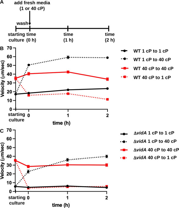
Fig 3.

Changes in swimming velocity upon changes in extracellular viscosity over time. (A) Outline of experimental procedures. WT C. jejuni and C. jejuni ΔvidA were grown overnight in MH broth alone (1 cP) or with methylcellulose (40 cP) for starting cultures. Cultures were washed, split, and then resuspended in fresh MH media alone or with methylcellulose (40 cP) and incubated as standing cultures in microaerobic conditions at 37°C. Swimming velocities of individual cells (n > 100) in starting cultures or at times 0, 1, and 2 h after introduction into fresh MH media at 1 or 40 cP were measured by video tracking under dark-field microscopy. (B and C) Swimming velocities of WT C. jejuni (B) and C. jejuni ΔvidA (C) over time. For panels B and C, average swimming velocities (±standard errors) of cultures at each time are shown. Starting cultures represent cells after overnight growth in MH at 1 or 40 cP. Measurements at time 0 h are taken immediately after resuspension of washed cells in fresh MH media alone (1 cP) or MH with methylcellulose for 40 cP. Black solid lines with circles indicate cultures grown overnight in 1-cP media and introduced into fresh media of the same viscosity, whereas solid red lines with squares indicate cultures grown overnight into 40-cP media and introduced into fresh media of the same viscosity. Dotted black lines with squares indicate cultures grown overnight in 1-cP media and then switched to 40-cP media, whereas dotted red lines indicate cultures grown overnight in 40-cP media and then switched to 1-cP media.
