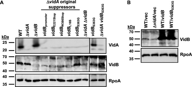
Fig 5.
VidA and VidB levels in WT C. jejuni and mutants. (A) Immunoblot analysis of VidA and VidB in whole-cell lysates of WT C. jejuni, ΔvidA, ΔvidA suppressor mutants with higher swimming velocities, and vidB mutants constructed in WT C. jejuni or ΔvidA. (B) Immunoblot analysis of VidB in whole-cell lysates of WT C. jejuni or ΔvidA containing vector alone or vector to overexpress vidB or vidBS363G. For panels Aand B, specific antiserum to VidA and VidB was used to detect each protein. Detection of RpoA serves as a control to ensure equal loading of proteins across strains. Note that a background band of ~50 kDa is observed around the same size of VidB in ΔvidB that hinders fully visualizing the absence of VidB in certain mutants.

