Figure 1.
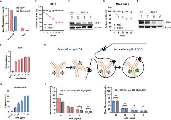
Antibody h128-3 induces LILRB4 internalization in monocytic AML cells. (A) Detection of LILRB4 expression on THP-1 and Mono-mac-6 cells by flow cytometry using an anti-LILRB4 rabbit mAb (R193) (n = 4). (B, D) Surface LILRB4 downregulation in THP-1 (B) and Mono-mac-6 (D) cells following treatment with h128-3 or control hIgG (n = 2). Cells were treated with 10 μg/ml of h128-3 or irrelevant hIgG. At different time points, cells were collected and surface LILRB4 was measured by flow cytometry using anti-LILRB4 rabbit mAb (R193) recognizing a different epitope than that targeted by h128-3. Surface LILRB4 was normalized to hIgG isotype control (100%). (C, E) Total LILRB4 measured by western blot. THP-1 (C) and Mono-mac-6 (E) cells were treated with h128-3, PBS or isotype control hIgG. On hours 24, 48 and 72, cells were collected and LILRB4 was measured by western blot using an anti-LILRB4 rabbit mAb recognizing a linear epitope (R8). (F, G) LILRB4 internalization in THP-1 (F) and Mono-mac-6 (G) cells induced by h128-3 with different concentrations (n = 4). Cells were treated with h128-3 and cultured at 37°C for 24 h before measurement of surface LILRB4 by flow cytometry. (H–J) Antibody internalization tested using a pH-dependent dye bound to antibody Fc that fluoresces in acidic intracellular environment (H). h128-3 and isotype control hIgG were conjugated with pH-dependent dye (pHAb) before addition to THP-1 (I) and Mono-mac-6 (J) cells at different concentrations (n = 4). Cells were cultured at 37°C for 24 h before fluorescence signal detection by flow cytometry.

