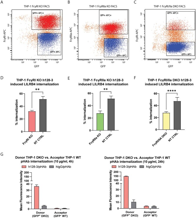
Figure 4.
FcγRIIa contributes to h128-3-induced LILRB4 internalization in FcγR high monocytic AML in a time- and FcγRI-dependent manner. (A–C) FcγRI (A) and FcγRIIa (B) were knocked out in THP-1 cells and FcγRI (C) was knocked out in THP-1 FcγRIIa KO cells by lentiviral particle transduction and positive selection of genes encoding Cas9-PuroR and sgRNA-eGFP or sgRNA-Bls or non-targeting control (NT CTRL) sgRNA-eGFP. Cas9 was doxycycline-induced to perform a double-stranded break of DNA encoding FcγRI or FcγRIIa at the sgRNA-guided sites. Cells were negatively selected by FACS after staining with APC-conjugated anti-FcγRI (10.1, Biolegend) and anti-FcγRIIa (2C3B11B8, Sino) mAbs. (D-F) h128-3-induced LILRB4 internalization in THP-1 NT CTRL cells relative to that in FcγRI KO (n = 4) (D) FcγRIIa KO (n = 4) (E) or FcγRI/FcγRIIa DKO cells (n = 4) (F). In brief, 2.5 × 10⁵ cells were treated with 5 μg/ml of antibodies at 37°C for 24 h before measurement of LILRB4 internalization by flow cytometry. (G) pHAb internalization in THP-1 FcγRI/FcγRIIa DKO (GFP−) donor cells or co-cultured THP-1 WT (GFP+) acceptor cells. Donor cells were opsonized with 10 μg/ml h128-3/pHAb or hIgG/pHAb and co-cultured 1:1 with acceptor cells for 4 or 24 h at 37°C before pHAb fluorescence in donor and acceptor cells was measured by flow cytometry and normalized to that of untreated co-cultured cells (n = 4).

