FIG. 5.

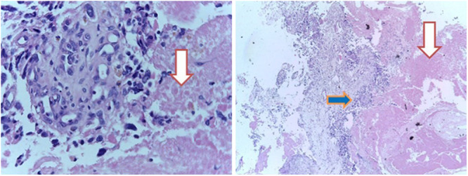
The histopathological appearance of the tumor under higher (left, original magnification ×20) and lower (right, original magnification ×4) magnifications showing extensive tumor necrosis (downward-facing arrows) seen in GBM. The right-facing blue arrow signifies a highly cellular sheet of hyperchromatic malignant glial cells with extensive mitosis.
