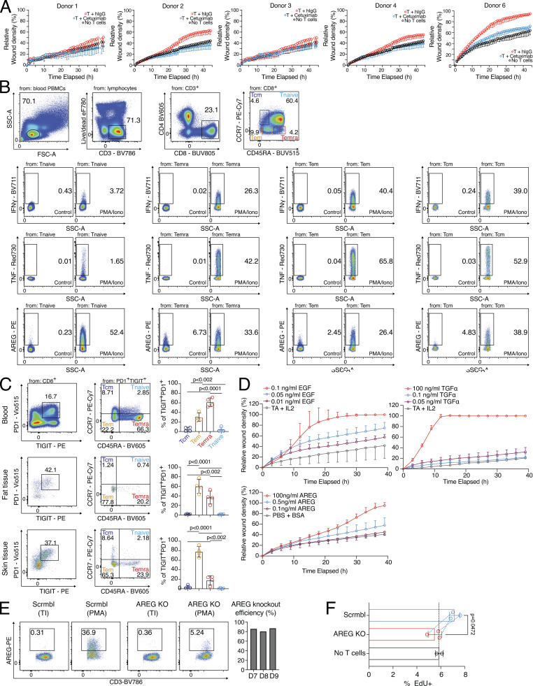
Figure S3.
Cytokine expression and wound healing potential of blood-derived CD8 T cells. (A) Wound healing assay with SNs of human blood-derived and in vitro activated and cocultured PD1+TIGIT+ CD8 T cells (T = T cells), treated with either human IgG or Cetuximab during wound healing assay; several donors. (B) Gating to identify Tnaive (CD45RA+CCR7+), Temra (CD45RA+CCR7−), Tem (CD45RA−CCR7−), and Tcm (CD45RA−CCR7+) in human blood, followed by intracellular flow cytometry to detect human AREG, TNF, and IFN-γ following 4 h incubation in the presence of PMA/ionomycin and transport inhibitors or transport inhibitor only. (C) Gating to identify Tnaive (CD45RA+CCR7+), Temra (CD45RA+CCR7−), Tem (CD45RA−CCR7−), and Tcm (CD45RA−CCR7+) in PD1+TIGIT+ CD8 T cells from human blood, fat, and skin tissue. Statistics to the right (one-way ANOVA, n = 4). (D) Wound healing assay with varying concentrations of recombinant human EGF, TGFα, or AREG. (E) AREG CRISPR KO efficiency. Left, exemplary flow cytometry plots depicting AREG expression; right, quantification. (F) EdU incorporation in HaCaT cells treated o/n with SN from scrmbl CD8 T cells, AREG knockout CD8 T cells or no T cell control (n = 3, Student’s t test). All data were derived from experiments with several independent donors.

