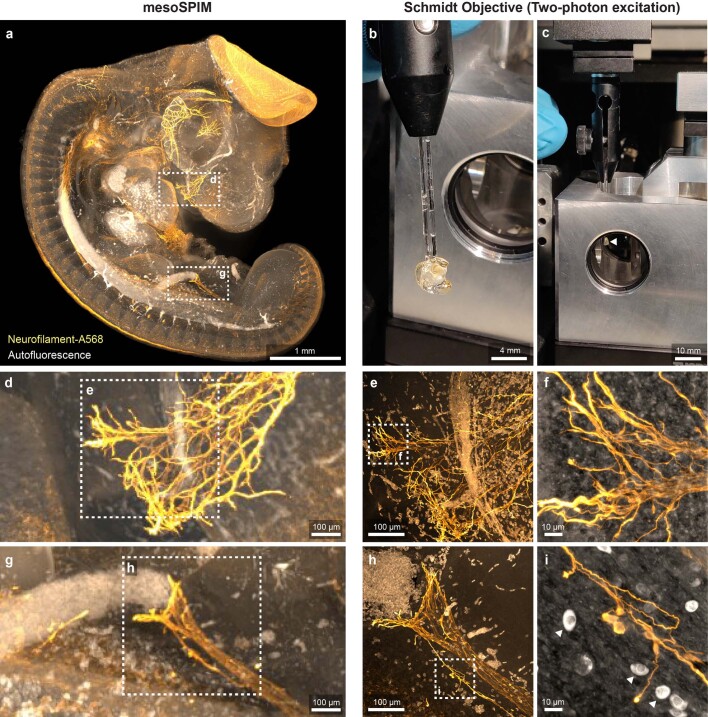
Extended Data Fig. 5. Correlative imaging of a 4-day old Chicken embryo with the mesoSPIM light-sheet microscope and the Schmidt objective.
A chicken embryo was stained for Neurofilament using Alexa 568 as a secondary antibody and cleared using BABB. a) Maximum projection of a single photon light-sheet (mesoSPIM) overview dataset of the sample. b) Sample (glued to a glass capillary) prior to insertion into the Schmidt objective. c) Sample (white arrow) inserted into the Schmidt objective. d) & g) Maximum projections (mesoSPIM dataset) of a region of interest chosen for high-resolution imaging. e) & h) Maximum projections of z-stacks taken at the highlighted locations in d) & g) using the Schmidt objective and two-photon excitation. f) & i) Maximum projections of stacks taken at higher zoom level showing individual axons. The white cells with strong autofluorescence are red blood cells which have not lost their nucleus yet at this developmental stage. The images are examples from one of two imaged chicken embryos.

