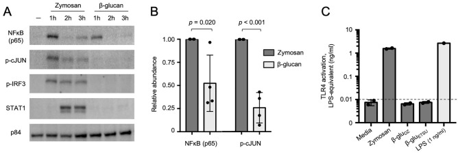
Figure 3.
Zymosan and β-glucans activate distinct signaling networks. (A) Immunoblot of nuclear extracts of monocytes stimulated with zymosan or ETSU β-glucan over a 3-h timecourse. Representative image from four replicates. (B) Quantification of nuclear NFκB (p65) and phospho-cJUN at 1 h after stimulation with indicated ligands, normalized to p84, aggregate of four replicates. (C) TLR4 activity measured by NFκB-induced SEAP activity in HEK-Blue TLR4 cells, quantified using LPS standard curve; dashed line represents limit of quantitation; zymosan 1 µg/ml, β-gluDZ and β-gluETSU 10 µg/ml.

