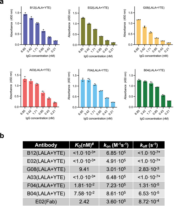
Fig. 2. IgG binding assessment.
a Dose-response ELISAs showing binding to myotoxin II of the clones as IgGs. Experiments were performed in triplicates (n = 3) and reported as means with error bars representing the SD and dots representing individual data points. b Affinity measurements of the six IgGs and one Fab determined in bio-layer interferometry experiments. *: The IgG was not observed to dissociate from myotoxin II, thereby resulting in the minimum value. #: Since six of the antibodies were measured as IgGs, the KD refers to functional affinity (avidity) for these. Source data are provided in a Source Data file.

