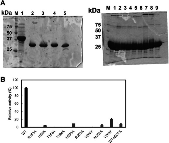
Fig. 5. Enzyme activity of site-directed mutants of LrhKduI.

(A) SDS-PAGE profile of the enzymes used in the assay. (left) M, marker; 1, S. agalactiae UGL; 2, LrhKduI WT; 3, LrhKduI R163A; 4, LrhKduI H200A; and 5, LrhKduD. (right) M, marker; 1, LrhKduI WT; 2, LrhKduI R163A; 3, LrhKduI I165A; 4, LrhKduI T184A; 5, LrhKduI T194A; 6, LrhKduI R203A; 7, LrhKduI Y207F; 8, LrhKduI M262A; and 9, LrhKduI Y269F. (B) Enzyme activity of LrhKduI mutants. WT activity was taken as 100 %. WT + EDTA indicates the LrhKduI WT enzyme in the presence of EDTA. Each data represents the average of triplicate individual experiments, except for H200A.
