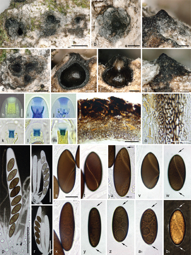
Fig. 18.

Spiririma gaudefroyi. a–b, d. Habit of ascomata on host surface in top view; c, g. erumpent ascomatal apices in side view; e–f. ascomata in vertical section; h–m. apical apparatuses in black Pelikan ink (h), diluted blue Pelikan ink (i–j), Melzer’s reagent (k–l) and Lugol’s solution (m); n. clypeus and upper peridium in vertical section, in chloral-lactophenol; o. lateral peridium in vertical section, in chloral-lactophenol; p. eight-spored ascus with paraphyses; q–r. mature and immature few-spored asci; s–b1. ascospores showing a helicoid germ slit (s–w, b1) and bipolar secondary appendages (arrows) (p–r, bl in diluted Indian ink; s–a1 in 1 % SDS) (a, c–e, s–w. WU-MYC 0044041 (epitype); b, f–r, x–b1. WU-MYC 0044043). — Scale bars: a–b, d = 500 μm; c, e–g = 200 μm; h–m = 5 μm; n, p–r = 50 μm; o = 20 μm; s–b1 = 10 μm.
