
Fig. 13.
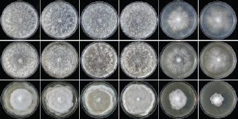
Colony morphology of Halophytophthora brevisporangia (BD662 and BD695), H. celeris (BD646 and BD885) and H. frigida (BD650 and BD655) (from left to right) after 7 d growth at 20 °C on sV8A, sCA and sPDA (from top to bottom).
Official websites use .gov
A
.gov website belongs to an official
government organization in the United States.
Secure .gov websites use HTTPS
A lock (
) or https:// means you've safely
connected to the .gov website. Share sensitive
information only on official, secure websites.
Colony morphology of Halophytophthora brevisporangia (BD662 and BD695), H. celeris (BD646 and BD885) and H. frigida (BD650 and BD655) (from left to right) after 7 d growth at 20 °C on sV8A, sCA and sPDA (from top to bottom).