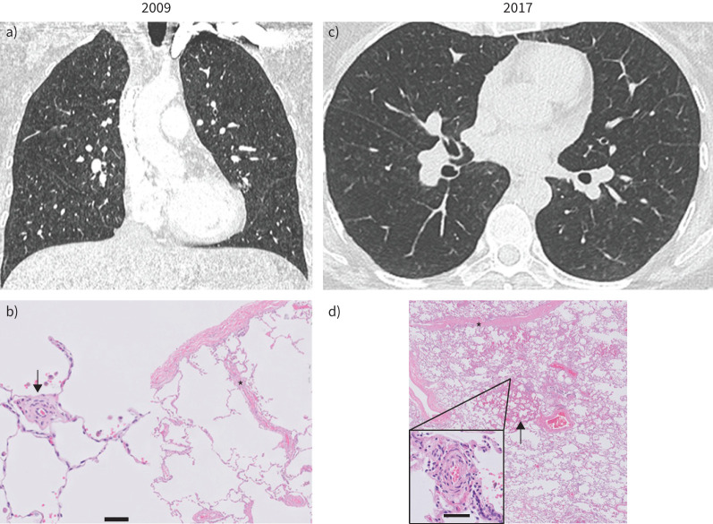
FIGURE 1.
Computed tomography and histopathological images of patient 1 at diagnosis (2009) (a and b) and at the time of lung transplantation (2017) (c and d). a) In 2009, chest computed tomography revealed mild nonspecific ground-glass opacities and interlobular septal thickening, without lymph node enlargement (not shown). c) In 2017, ground-glass opacities and interlobular septal thickening are more pronounced. b) Lung histology in 2009 (surgical biopsy, haematoxylin and eosin staining) and d) in 2017 (lung transplant, haematoxylin and eosin–saffron staining) are shown. In b), pulmonary microcirculation (scale bar = 50 µm) shows extensive thickening of the intima (arrow) with subtle venous remodelling (asterisk). d) Subtotal obliteration of septal veins is confirmed in the explanted lung in 2017 (asterisk) with patchy capillary congestion and proliferation (arrow) and more extensive remodelling of the microcirculation (zoom-in panel; scale bar = 50 µm).

