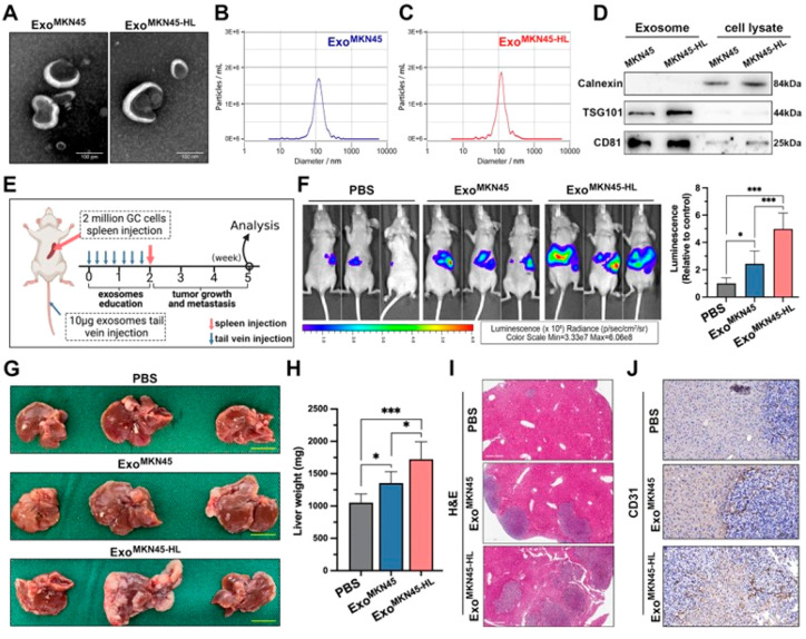
Figure 4.
Exosomes associated with liver metastasis in gastric cancer (GC) show distinct characteristics. (A) Transmission electron microscopy (TEM) image of exosomes derived from MKN45 and MKN45-HL cells and (B, C) size distribution analysis of purified exosomes from MKN45 and MKN45-HL cells using NanoSight. (D) Western blot assessment of exosome markers (TSG101 and CD81) in exosomes and lysates from MKN45 and MKN45-HL cells. (E) Diagram illustrating the process of establishing the exosome-informed GC-LM model. (F) Representative in vivo imaging system (IVIS) outcomes in mice injected with luciferase-tagged MKN45 cells into the spleen after being exposed to PBS, MKN45 exosomes, or MKN45-HL exosomes. (G–I) Impact of GC-derived exosomes on liver metastasis in mice, featuring images of liver metastasis, liver weight, and H&E staining. (J) Representative CD31 immunohistochemical staining images of liver metastasis tissues from exosome-educated mice (Adapted with permission from ref (118). The copyright is licensed under a Creative Commons Attribution 4.0 International License 2022, .J Exp. Clin. Cancer Res., Springer Nature).

