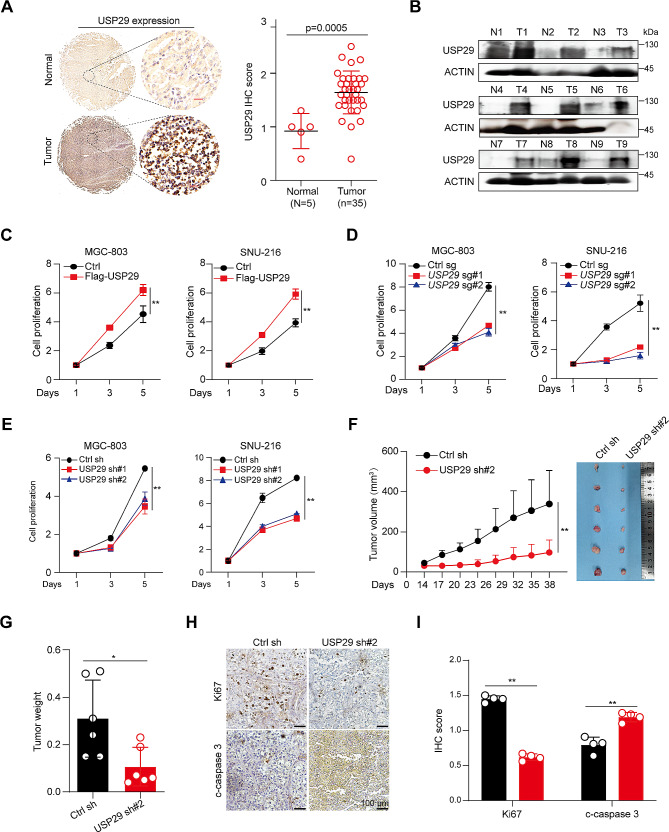
Fig. 1.
Overexpression and an oncogenic role of USP29 in gastric cancers. (A) Left: Representative immunohistological images of USP29 in 5 normal gastric tissues and 35 tumors. Scale bar: 20 μm; right: Quantifications of the immunohistochemistry in the left are shown. (B) USP29 protein expression in 9 gastric tumors and paired non-cancerous tissues analyzed using Western blot. (C) Cell proliferation of MGC-803 and SNU-216 cells with or without USP29 overexpression. (D) Cell proliferation of MGC-803 and SNU-216 cells with or without USP29 depletion by specific sgRNAs. (E) Cell proliferation of MGC-803 and SNU-216 cells with or without USP29 knockdown by specific shRNAs. Data shown were obtained from mean ± SD of technical triplicates (C-E). (F) MGC-803 xenograft tumor growth curves and tumor image (n = 6). (G) Tumor weights of MGC-803 xenografts (n = 6). Data shown were obtained from mean ± SD of technical triplicates (F, G). (H) Representative immunohistological images of Ki-67 and cleaved-caspase-3 in MGC-803 xenograft tumors. Scale bar: 100 μm. (I) Quantifications of the immunohistochemistry in H. **p < 0.01

